

 PDF
PDF ePub
ePub Citation
 Print
Print


Introduction
Hydatid disease or echinococcosis, is mainly caused by the larval stage of taenia Echinococcus granulosus1. It is prevalent in sheep raising areas in the Mediterranean region, South America, Australia, New Zealand, Russia and China2. Recently, the disease has been emerging in Europe and North America, due to increased travel and immigration1. Humans are accidental intermediary hosts in the biological life cycle of taenia Echinococcus granulosus. The taenia's eggs frequently infest by direct contact with dogs and sheep3. After enteric digestion, the liver and the lungs are most commonly affected via the bloodstream in adults3. Pleural involvement of hydatid disease can occur, and usually follows the rupture of a pulmonary or hepatic hydatid cyst into the pleural space4. However, primary pleural involvement by a slowly enlarging cyst is very rare. Herein, a case of primary pleural hydatid cyst infestation was diagnosed by video-assisted thoracoscopic surgery in a patient that presented with a slow growing pleural nodule.
Case Report
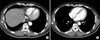
A 50-year-old man without any significant past medical history presented with a 2 cm nodular lesion in the right lower lateral lung field noted on a plain chest radiography. The nodule was detected during the evaluation of diabetes mellitus when the patient presented with diabetic retinopathy. The patient had denied any exposure to asbestos, overseas travel and respiratory symptoms including cough, sputum, dyspnea, hemoptysis, fever and chest pain. There was no history of ingestion of freshwater crabs, or raw beef. Physical examination revealed normal breath sounds and no focal chest wall tenderness. Laboratory test results showed a high HbA1c of 9.5%, but no leukocytosis or eosinophilia was observed. High sensitivity C-reactive protein, carcinoembryonic antigen (CEA), CA 19-9, and AFP were all within normal range. The test for human immunodeficiency virus (HIV) was negative. Abdominal sonography showed only mild fatty liver without any cysts or masses in the liver or spleen. A plain chest radiography showed a 2 cm nodular lesion in the right lower lateral lung field. The chest computed tomography (CT) scan showed a 2.2×1.9 cm well demarcated pleural based nodule between the lateral arc of the right seventh and eighth ribs with internal fat density (Figure 1A), suggesting the possibility of a benign pleural tumor such as a lipoma. On the follow up chest CT scan, three months later, the size of the pleural nodule increased about 4 mm and there was still an internal fat density (Figure 1B). A liposarcoma was suspected and video assisted thoracoscopic surgical excision was performed. At the 8th intercostal space, round cystic nodule was found with adhering to pleura. A 2.7×2.5×0.7 cm sized pleural cystic nodule was extirpated. Histopathological examination showed a hydatid cyst (Echinococcosis) between the visceral and parietal pleura with fibrosis (Figure 2). The patient was discharged five days after the operation without complications. There was no evidence of disease recurrence by chest CT scan during the 18 months of follow up.
Discussion
Adult Echinococcus granulosus lives in the intestinal tract of infested dogs. Its eggs are excreted in the dog's feces and swallowed by intermediate hosts, such as sheep, cattle, goats, and humans. Once a human has been infested with the taenia eggs, gastric and enteric digestion facilitates the release of larvae, which penetrate the intestinal wall until they reach a small vessel system. Passing through the bloodstream, they arrive at the organ where they can settle and transform into small cysts that increase in size by 2 to 3 cm per year5. The usual locations are the liver and lungs; intrathoracic but extrapulmonary locations like the pleura, diaphragm, mediastinum, pericardium, and chest wall are uncommon6. Pleural hydatid cysts can develop chiefly as a result of liver or lung cyst rupture into the pleural space with complications of pneumothorax, pleural effusion or empyema1. However, a hydatid cyst located primarily in the pleural space, as observed in this case, is very rare. Only about 15% of larvae succeed in passing through the hepatic and pulmonary capillary barrier to reach the systemic circulation7. Hydatid cysts consequently can be found in any tissue but more vascularized tissues have a greater chance of implantation. Because pleural membranes have only small blood capillary networks, it is difficult for the cysts to reach the pleural space through the bloodstream6. It is uncommon even in an endemic country that hydatid disease directly invading the pleura without hepatic or pulmonary involvement. This is the first report of primary pleural hydatid disease in a non-endemic country.
About 90% of patients with hydatid disease present with a variety of symptoms including cough, chest pain, hemoptysis, malaise, fever and even expectoration of cystic materials2. The patient presented here was asymptomatic and diagnosed incidentally on a plain chest radiography during the work up for diabetes mellitus. The diagnosis of hydatid disease still depends on radiography8. Hydatid cysts in the liver or lungs are diagnosed easily but intrathoracic extrapulmonary cases may have atypical radiological presentation6. In this case, the lesion was presumed to be a neoplasm of the pleura. Laboratory tests can complement the clinical and radiological investigations. Blood testing is usually negative for eosinophilia in cases with intrathoracic hydatid disease8. Immunological tests such as IgG ELISA, indirect hemagglutination assay, and the hydatid antigen dot immunobinding assay can be helpful, but the sensitivity is only around 60%9. A combination of two or more tests and radiology imaging should be used for an accurate diagnosis.
Surgery is the treatment of choice; cystectomy and capitonnage are the standard procedures10. During surgery, spillage of hydatic fluid must be rigorously avoided because of the risk of anaphylaxis and disease recurrence 11. Postoperative morbidity and mortality are low, from 1 to 2%8. In this case, the hydatid cyst was completely removed without any spillage of hydatic fluid. There was no recurrence detected over 18 months of follow up by chest CT scan.
International population movement is an integral component of the globalization of infectious diseases including viruses12, bacteria13, and parasites14. Some parasitic diseases are slowly spreading globally with global warming and are a public health concern14. The patient presented here lives in Ansan city, for 12 years and has no history of overseas travel. Ansan city is an industrial city of 750,000 residents with more than 1,000 factories and more than 35,000 foreign workers who came from China (25,000 persons), Vietnam (2,500 persons), Philippine (1,500 persons), Russia (700 persons), Iran (50 persons), and Israel (10 persons) (http://stat.iansan.net). Among them, a considerable number came from hydatid disease endemic countries such as China, Russia, Iran, and Israel. The patient works on construction along with many foreign colleagues, and has occasionally contacted with wild dogs in the field of construction. It was assumed that the infested foreign workers entered Korea. Their sputum containing hydatid cysts were expectorated and accidentally ingested by dogs. The contaminated food or water with dog's stool was eaten by the patient. In Korea, the prevalence survey of parasites in dog revealed no echinococcus15. Echinococcus seemed to be not naturalized in Korea. Although hydatid disease is rare in developed countries, greater population mobility and migration may increase the frequency of this disease. In the differential diagnosis of pleural disease, pleural hydatid disease should be included even if there is no overseas travel history.

 XML Download