

 PDF
PDF ePub
ePub Citation
 Print
Print


Introduction
The term 'tuberculous-destroyed lung (TDL)' is usually used to describe the destructive lung parenchymal changes due to sequelae of pulmonary tuberculosis, which occur over years, and cause chronic airway obstruction as well as restrictive change. In South Korea, tuberculosis (TB) burden is intermediate (88/100,000) and complications from TB are still an important pulmonary disorder1, but clinical manifestations of TDL can be similar to those of chronic obstructive pulmonary disease (COPD) with manifestations of dyspnea due to airway obstruction. Furthermore, TDL may be a common cause of acute exacerbation with respiratory failure in South Korea2, but there are few predictive factors to suggest differences in the prognosis between patients with TDL and those with COPD when patients with dyspnea, caused by aggravation of airway obstruction, are admitted to intensive care unit (ICU) until now.
Chronic bronchitis or bronchilolitis and emphysema can occur as complications of pulmonary TB, and the degree of airway obstruction changes in subjects treated for TB; it increases with age, the number of cigarettes smoked, and the extent of the initial TB disease3. However, it is difficult to distinguish patients with airway obstruction due to TDL from patients with pure COPD on initial presentation with dyspnea. The suggested mechanism of airway obstruction in TDL is a sequela of tuberculosis, due to granulomatous changes in the bronchial wall, extrinsic pressure from enlarged peribronchial lymph nodes, or residual fibrotic endobronchial stenosis4, while those of COPD are chronic inflammatory disease of the lungs, mostly in response to cigarette smoking5.
TDL patients with acute respiratory failure and positive AFB smears have higher mortality rates than those with negative AFB6. Furthermore, patients with pulmonary TB presenting with dissemination are more likely to present with acute respiratory failure requiring mechanical ventilation and to have a worse prognosis than patients with localized TB pneumonia7. However, a radiological evaluation, including high-resolution computed tomography (HRCT), does not provide a definite clue to the differential diagnosis from bacterial pneumonia, so an early diagnosis can be delayed.
In cases of COPD, mortality is related to the frequency of severe exacerbations requiring hospital care, and the presence of acute physiology and chronic health evaluation (APACHE) II-associated comorbidity and the need for mechanical ventilation ≥72 hours are independent predictors of poor outcome8.
Although several published studies have focused on patients with active pulmonary TB and respiratory failure, few studies have attempted to identify the difference between patients with TDL and COPD who have respiratory failure symptoms. Thus, the aim of this study was to identify differences in the clinical features between patients with TDL and COPD who were admitted to ICU due to dyspnea.
Materials and Methods
We reviewed the medical records of patients with TDL and a TB history, who had an obstructive pattern on a pulmonary function test and patients with COPD without a TB history who had an obstructive pattern on a pulmonary function test. They were all admitted to the ICU of a Busan Paik University-Affiliated Hospital via the emergency room due to dyspnea between October 2004 and May 2009. The patients with accumulated available pulmonary function test results with less than 70% of forced expiratory volume in 1 second/forced vital capacity (FEV1/FVC) were selected (best value closest to admission day), and patients who had co-existing pulmonary or cardiovascular diseases were excluded.
Initially 38 patients with TDL and 39 with COPD were selected according the above criteria, but patients with underlying diseases such as malignancy, heart failure, and chronic renal failure as well as previously unconfirmed patients with COPD were excluded from the final analysis. Also, patients who had a comorbidity other than an airway obstruction, such as a pneumothorax, massive hemoptysis, and pleural effusion, which could confound the dyspnea caused by airway obstruction, were excluded.
Sixteen patients with TDL and 16 patients with COPD were finally enrolled, and we analyzed demographic data, pulmonary function test results, arterial blood gases, blood chemistry, radiological characteristics, APACHE II sores, combined medical problems on admission, total ventilation time, and final outcome in the ICU.
1. Definition of TDL and COPD
We defined patients with TDL as those presenting with lung function insufficiency due to previous pulmonary TB, parenchymal damage to more than one lung lobe, no recent evidence of active TB, less than 20 pack-years smoking history, and no current smoking habit9. For COPD, we followed the definition of the Global Initiative for Chronic Obstructive Lung Disease10.
2. Statistical analysis
The SPSS version 12.0 (SPSS Inc., Chicago, IL, USA) software was used for all statistical analyses. The chi-squared test or Fisher's exact test was used for categorical variables. The student t-test for pulmonary function tests and the Mann-Whitney test for other continuous variables were used. Statistical significance was defined as a p-value <0.05.
Results
1. Clinical characteristics of the patients with TDL and COPD
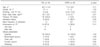
Thirty-two subjects were finally analyzed in the study (Table 1). The mean ages of the patients with TDL and COPD were 63.7 and 71.2 years, respectively. The mean body mass index (BMI) was 19.7 and 21.8 kg/m2, respectively, showing that the patients were in a malnourished state.
All patients with COPD had a smoking history of more than 20 pack-years, while none of the patients with TDL did. No difference was observed between the two groups for compliance with regular follow-up in the outpatient clinic before admission. Patients with TDL had no more frequent admission histories than patients with COPD (50.0% vs. 68.7%), and patients with TDL were admitted to hospital as frequently as patients with COPD (mean admission frequency, 1.4 times vs. 3.6 times, respectively).
Emphysematous changes on a chest CT were seen in 14 of 16 patients (87.5%) in the TDL group and in 9 of 16 patients (56.3%) in the COPD group all with multi-lobe involvement. The frequency of blood-tinged sputum and chest pain tended to be higher in the patients with TDL than in those with COPD, but the difference was not significant.
2. Differences between patients with TDL and patients with COPD
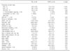
As shown in Table 2, the predicted FEV1/FVC (%) was similar in the two groups (55.0% vs. 55.2%), but the mean FVC predicted value was significantly lower in patients with TDL than in those with COPD (50.4% vs. 71.9%; p<0.01), and FEV1 values were significantly lower in patients with TDL than in those with COPD (39.1% vs. 58.4%; p<0.01). When a positive bronchodilator response which was officially defined as an absolute change in FEV1 of more than 200 mL and a percentage of initial FEV1 of more than 12% was applied11, the positive rates were 6.3% in patients with TDL and 18.8% in those with COPD, respectively.
Consolidation was more frequently found on X-ray in patients with TDL than in those with COPD (68.8% vs. 31.3%; p=0.03), and 6 of 16 patients (37.5%) with TDL were confirmed as recurrent pulmonary TB with a positive AFB smear and culture results, which was a significantly higher rate than patients with COPD (37.5% vs. 0%; p=0.02).
No significant difference was observed for PaO2/FiO2 ratio between patients with TDL and COPD. The PaCO2 tended to be higher in patients with TDL (54.9±25.4) than in those with COPD (44.7±21.1), but the difference was not significant. Also, APACHE II scores were not different between two groups. A ventilator was used in 8 of 16 patients (50.0%) in the TDL group, and 10 of 16 patients (62.5%) in the COPD group, among which non invasive ventilation was used in 1 COPD patient.
Four patients in the TDL group underwent a tracheostomy, and the procedure was performed at an average of 10.5 days after intubation. ICU stay days and total ventilation time were longer in the TDL group than the COPD group without statistical significance. Mean arterial pressures were not different between the TDL group and the COPD group (85.6±29.6 mm Hg vs. 76.2±18.5 mm Hg), but blood urea nitrogen was significantly lower in the TDL group than in the COPD group (27.8±22.6 mg/dL vs. 51.5±44.2 mg/dL; p=0.03).
The main cause of dyspnea in patients with TDL was thought to be an exacerbation of chronic airway obstruction, aggravated by associated respiratory infection, mostly manifesting as a pneumonic consolidation on chest X-ray. Among patients with TDL, wheezing sounds were audible among 6 of 16 patients (37.5%), and steroids were used in 10 of 16 (62.5%) patients at initial presentation. The survival rate was not different between the two groups (81.3% vs. 87.5%), and the cause of mortality of TDL patients (n=3) and COPD patients (n=2) were multi organ failure due to septic shock alike.
Discussion
The pulmonary function test results of patients with TDL were more severely diminished than those of patients with COPD. In addition, the patients with TDL had TB pneumonia more frequenctly and more tracheostomies than patients with COPD in ICU setting. This study identified differences between patients with TDL and COPD upon ICU admission excluding patients with underlying disease such as heart failure and renal failure, which is different from previous studies that described lung functions of patients in out-patients setting9.
Loss of lung volume in patients with TDL can be explained in view of the destructive and fibrosing properties of pulmonary TB causing pulmonary overdistention3. The obstructive lung disease might be attributable to TB involvement of the bronchial tree, with endobronchial inflammation12, bronchiectatic lesions caused by TB13, or emphysematous parenchymal changes by traction due to extensive scarring14,15. In our study, all of 16 patients with TDL actually had emphysema with TB scar and fibrotic changes instead of pure emphysema on HRCT scan. Furthermore, the lower positive bronchodilator response rates in patients with TDL may be due to the irreversibility of anatomical airway constriction in TDL. Therefore, the stay time in the ICU tended to be longer in patients with TDL than in patients with COPD, because weaning was more difficult in patients with TDL. In our study, a tracheostomy had been performed on four patients of TDL; decision for tracheostomy after day 10 of mechanical ventilation was made by full-time critical care physicians who followed the weaning protocol16. Increased elastic and restrictive work loads must have contributed to difficult weaning in the patients with TDL as with the patients with COPD, but the main mechanism is unknown17. Additionally, when we analyzed the patients with similar PFT values categorized by 30~80% of the FEV1 value, there were no additional clinical differences between TDL group (n=11) and COPD group (n=13) except platlet count (255.4±89.2×10⁹/L versus 187.1±83.0×10⁹/L, p=0.04).
In our study, the main pathogens for pneumonic consolidation were S. pneumoniae, P. aerosinosa, and K. pneumonia without difference between the TDL group and COPD group, but as many as 6 of 16 TDL patients were confirmed with recurrent pulmonary TB. Because TB pneumonia may frequently be indistinguishable from bacterial pneumonia, the diagnosis is generally made later. TB pneumonia more commonly occurs in older patients and in immunocompromised individuals, such as those with diabetes mellitus, cancer or infection with human immunodeficiency virus18,19. In our results, consolidation was predominantly observed in anatomically distorted areas induced by extensive TB scaring, especially in patients with TDL. Thus, it must be remembered that in patients with TDL and airflow obstruction, manifesting dyspnea, for whom intensive cares are needed, aggressive diagnostic strategy should be considered to exclude TB reactivation, especially in cases with consolidation.
Frame et al. reported that the mortality rate for patients with TB and respiratory failure was 81%, and that of patients with TB requiring intensive care was 67%20. But, in our study, none of the six recurrent patients with TB in TDL requiring intensive care died. Compared with a previous study21, the reason for the lower mortality rate of these patients could be due to the lack of a smoking history, no underlying heart disease, and a smaller extent of lung destruction in our patients.
To appreciate our results correctly, we should consider several limitations of the study. First, the number of cases involved was small. Second, co-existing asthma could not be completely excluded in either group. Lastly, not all patients were under ventilator care in the ICU. Therefore, prospective and larger studies are needed to clarify the main mechanism and the characteristics of dyspnea exacerbation in patients with TDL and COPD.
In conclusion, TB pneumonia must be ruled out in patients with TDL, presenting with dyspnea exacerbation due to airflow obstruction, and more severe restriction and obstruction of pulmonary function, with a tendency for a longer ICU stay, are evident in patients with TDL versus those with COPD.
 XML Download