

 PDF
PDF ePub
ePub Citation
 Print
Print


Introduction
Lung cancer is known as the most fatal cancer, and the incidence is increasing1,2. Most patients with lung cancer are inoperable at the time of diagnosis, and the main therapeutic strategy is chemotherapy. Thus, there is growing interest about surrogate biomarkers to predict responses of chemotherapy to avoid serious side effects.
The presence of DNA and RNA in serum or plasma of cancer patients has been recognized since the 1970s. In 1977, useful application of circulating nucleic acid in cancer diagnosis and treatment was proved. The circulating DNA level was high in the serum of cancer patients, and it decreased when tumor responded to radiotherapy3.
Tumor-derived RNA has been detected in plasma or serum of cancer patients with nasopharyngeal carcinoma4 and melanoma5 using reverse transcriptase (RT) PCR. In addition, tumor-derived RNA may serve as a potential biomarker for non-invasive cancer monitoring. Cytokeratin 19 (CK19) and mammaglobin mRNA in the plasma of breast cancer patients is associated with a poor prognosis6.
While the quantification of plasma DNA has been proposed as a diagnostic tool for cancer7, quantitative analysis of circulating cell-free RNA (cRNA) has not been fully investigated. Moreover, the usefulness of total cRNA as a predictive biomarker has not been unknown. Recently, in vitro data with cancer cell lines showed that the level of circulating cRNA decreased immediately after exposure to anti-cancer drugs, suggesting that the metabolic rate of cells affects the cRNA level, and quantification of cRNA may be used as a potential biomarker for monitoring the response to chemotherapy8.
The purpose of this study was to determine whether the change in cRNA level before and after chemotherapy can be used as a biomarker to predict the response to chemotherapy in patients with lung cancer.
Materials and Methods
1. Patients and collection of serum samples
We evaluated 32 consecutive patients with pathologically-confirmed lung cancer who received care in the Pulmonology Department of Dong-A University Medical Center between June and December 2009. The eligibility criteria for the study were as follows: >18 years of age; bi-dimensionally measurable lesions; and scheduled to undergo chemotherapy. All participants gave written informed consent to participate in the study and the trial was approved by the Institutional Review Board. Individuals were excluded for the following reasons: received chemotherapy or radiotherapy within 4 weeks before enrollment; and conditions that influence serum concentration of nucleic acids, such as diabetes, autoimmune diseases, burns, severe trauma, and brain infarctions.
Blood samples were obtained from eligible individuals to measure cell-free RNA concentration within 1 week before chemotherapy and after 2 cycles of chemotherapy. Blood samples containing 0.1 volume of 3.8% citric acid were centrifuged at 3,500 rpm at 4℃, and then the separated serum samples were stored at -80℃ until assayed.
2. Clinical responses
Patients underwent assessment of treatment responses after two cycles of chemotherapy using computed tomography (CT) scans, and classified into complete response (CR), partial response (PR), stable disease (SD), and progressive disease (PD) according to the Response Evaluation Criteria in Solid Tumors9. All patients were divided into two groups according to the response to chemotherapy. The response group included CR and PR. Patients with SD, PD were regarded as the non-response group.
3. Detection of cell-free RNA in the serum
Total RNA was isolated from the serum by a RNAspin Mini RNA isolation kit (GE Healthcare, Uppsala, Sweden), according to the instructions of the manufacturer. The total yield of RNA isolated from one aliquot of serum was reverse transcribed with a mixed solution of 2×Reverse Transcription Master Mix by cDNA Reverse Transcription Kits (Applied Biosystems, Foster City, CA, USA), and the amount of cDNA corresponding to 75µL of serum was used for each RT-PCR experiment. A real-time quantitative RT-PCR assay (QRT-PCR, TaqMan format) was used for transcript quantification of the glyceraldehyde-3-phosphate dehydrogenase (GAPDH) gene. QRT-PCR was set up in a reaction volume of 25µL (4.5 mM MgCl2, 0.75 U TaqGold, initial denaturation [2 minutes at 50℃ and 10 minutes at 95℃], denaturation [40 cycles for 15 seconds at 95℃], and annealing and extension [1 minute at 60℃]). The primers were as follows: forward, 5'-GAA GGT GAA GGT CGG AGT C-3'; reverse, 5'-GAA GAT GGG ATT TC-3'; and probe, 5'-FAM-CAA GCT TCC CGT TCT CAG CC-3'. Amplification data were collected and analyzed with an ABI Prism 7000 Sequence Detector (Applied Biosystems). Each sample was analysed in duplicate, and multiple negative water blanks were included in every analysis. For calibration purposes the GAPDH amplicon was cloned into a plasmid and serially diluted (106-100 copies per tube).
4. Statistical methods
Continuous variables are presented as the mean±standard deviation. Categorical data are reported as frequencies. Differences in continuous variable were compared with the Mann-Whitney test or Wilcoxon signed-rank test. Dichotomous variables were compared by Fisher's exact test. The significance level was defined as a p<0.05. All analyses were performed with SPSS version 18.0 (SPSS Inc., Chicago, IL, USA).
Results
1. Patient characteristics
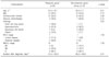
The mean age was 61.4 years, and 23 patients (69.7%) were men. Sixty-nine percent of the participants were former or current smokers. The histologic subtypes were as follows: 15 patients (45.5%), adenocarcinoma; 6 patients (18.2%), squamous cell carcinoma; 1 patient (3.0%), large cell carcinoma; 2 patients (6.1%), indeterminate non-small cell lung cancer (NSCLC); and 8 patients (24.2%), small cell lung cancer (SCLC). Twenty-one persons (65.6%) had no prior treatment, and received first-line chemotherapy. Most of patients in this study were enrolled within 3 months since pathologic diagnosis (n=28, 87.5%). The clinical characteristics were not different between the response and non-response groups (Table 1).
2. Serum total cell-free RNA expressed as the Ct GAPDH level
The clinical response of participants was as follows: 1 patient (3.1%), CR; 18 patients (56.3%), PR; 3 patients (9.4%), SD; and 10 patients (31.3%), PD. Thus, 19 patients (59.4%) were grouped as the response group according to the study protocol. It was possible to isolate intact RNA from all of the patients. The pre-chemotherapy cRNA expressed as Ct GAPDH level was not different between the response and non-response groups (response group 41.36±1.72 vs. non-response group 41.33±1.54; p=0.78). The post-chemotherapy Ct GAPDH level showed no difference between two groups either (response group 39.92±1.81 vs. non-response group 40.41±1.47; p=0.40). The increasing or decreasing trend of cRNA after chemotherapy did not showed a significant relationship with clinical responses. Patients who showed increased tendency of cRNA level after chemotherapy were sixty-five percent of response group and thirty-five percent of non-response group (p=0.43) (Table 2). We also could not find a correlation between cRNA level and clinical responses in subgroup analysis according to histologic subtypes, NSCLC and SCLC.
Discussion
The purpose of the current study was to determine the relationship between quantification of serum total cRNA level and clinical response to chemotherapy in patients with lung cancer. Though we could not demonstrate a significant relationship, this is the first report to investigate quantification of serum cRNA as a predictive biomarker for the response to chemotherapy in patients with lung cancer. It seems that quantification of total serum cRNA may not to be a useful biomarker for predicting a response of chemotherapy in patients with lung cancer.
The recent interest in serum/plasma nucleic acids has created many new promising possibilities for the non-invasive detection and monitoring of a variety of conditions10. It is also known that in cancer patients, a portion of these free nucleic acids are released from tumor cells. After the first demonstration of detectable tumor-associated DNA sequences in the plasma of cancer patients by Stroun et al. in 198911, different forms of tumor-derived circulating DNA were detected in patients with various types of cancers12-14. Both genomic and mitochondrial DNA quantification in the circulation have been extensively evaluated as diagnostic and prognostic tool to monitor cancer management. Cell-free DNA bearing the same genetic and epigenetic changes as the tumor tissues were detectable in plasma / serum of cancer patients12-14, suggesting tumor-specific DNA markers in blood can be useful for minimally invasive diagnostic tests.
Like circulating DNA, detection of tumor-derived RNA in plasma/serum is also a promising approach in cancer management. As circulating RNase is present in the blood of cancer patients, as well as in healthy individuals15, serum free RNA was expected to be rapidly destroyed in the blood. However, cell-free Epstein-Barr virus-associated RNA in the plasma of nasopharyngeal cancer patients could be detected by RT-PCR, similar methodology as for DNA analysis4. At the same time, tyrosinase mRNA in malignant melanoma patients was also demonstrated5. These findings are consistent with the idea that there is extracellular RNase protected tumor-derived RNA in the circulation. Based on that idea, there have been many trials to use circulating RNA for the diagnosis of tumors. Recently, plasma hnRNP B1 mRNA was reported as a useful non-invasive marker for the detection of lung cancer16.
Although the blood cell-free nucleic acid level is high in patients with cancer, there are many other conditions in which the level of cell-free nucleic acid level is increased, such as stroke, burns, severe trauma, myocardial infarction, and autoimmune diseases17-21. We excluded patients with those diseases.
The quantification of free circulating total DNA in the serum of cancer patients was proposed as a non-invasive diagnostic tool, because the increased level was demonstrated in patients with lung cancer compared to the matched benign control roup7. The plasma RNA level is known to be increased in many benign and malignant lung diseases22. Although the origins and clinical parameters for determining the quantity of total cRNA are still being identified, quantification of the total amount of c RNA might be a surrogate marker of the presence of malignancy like DNA21. However, recent investigation failed to demonstrate more increased level of total amount of RNA in blood of patients with lung cancer compared to patients with benign lung disease23. In addition, though other investigators observed a correlation between the increased amount of circulating tumor-related β-catenin mRNA and the presence of colorectal tumor, they did not find those correlation with the concentration of GAPDH mRNA, house-keeping gene, representing total amount of cRNA24. So further studies comparing patients with lung cancer and healthy control or patients with benign lung diseases are needed to prove diagnostic role of serum total cRNA.
It is also uncertain whether or not serum total cRNA can be used as a predictive biomarker of lung cancer treatment. In an in vitro study, investigators demonstrated that both hypoxia and a rapid metabolic rate are able to induce an increase in total cRNA8. Such is a promising result for quantification of total cRNA as a potential predictive biomarker of chemotherapy in patients with cancer. Our study was conducted to determine if quantification of total cRNA can be used in patients with lung cancer. However, the trend of level of cRNA did not reflect clinical response. Though it was not statistically significant, many patients of response group showed elevated level of cRNA after chemotherapy. Because the knowledge on the source of cell free RNA and more importantly the mechanism remains unanswered, it seems that the role of quantification of total cRNA in blood as a predictive biomarker in patients with cancer is still unknown.
The limitation of our study was small sample size. Furthermore, there is lack of universally accepted validated or standardized methods measuring amount of RNA. Although we examined the relationship between cRNA and the clinical response after chemotherapy according to histologic type, previous chemotherapy, and smoking status, there were no significant results.
In conclusion, the quantitative analysis of total cRNA in serum may not be useful in predicting the tumor response to chemotherapy in patients with lung cancer. To validate this result, additional studies with a larger sample size and more standardized and validated methods are required.

 XML Download