

 PDF
PDF ePub
ePub Citation
 Print
Print


Abstract
We report a case of Mycobacterium intracellulare pulmonary infection presenting as a solitary pulmonary nodule (SPN). A 35-year-old male was admitted due to a SPN in the right upper lobe which was detected on the chest radiography being examed due to recurrent cough for 1 year. The computed tomography (CT) revealed a spiculated nodule containing air-bronchogram, which was suspicious of malignancy. We performed transbronchial biopsy and the pathology showed granulomatous inflammation with caseous necrosis. Under the presumptive diagnosis of pulmonary tuberculosis, we started anti-tuberculous medication including isoniazid, rifampin, ethambutol, and pyrazinamide. In one month, however, the sputum culture was positive for Mycobacterium intracellulare. The follow-up chest CT showed slight aggravation of the previous lesions. Under the final diagnosis of Mycobacterium intracellulare pulmonary infection presenting as a solitary pulmonary nodule, we changed the regimen to rifampin, ethambutol, and clarithromycin. The follow-up chest CT after the completion of treatment, revealed resolution of the previous lesions.
Figures and Tables
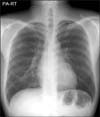
Figure 2
The initial chest CT shows a spiculated nodule with airbronchogram in the right upper lobe.
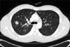
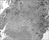
Figure 3
The histologic finding of the transbronchial biopsy specimen shows chronic granulomatous inflammation with caseous necrosis (H&E stain, ×100).


References
1. Griffith DE, Aksamit T, Brown-Elliott BA, Catanzaro A, Daley C, Gordin F, et al. An official ATS/IDSA statement: diagnosis, treatment, and prevention of nontuberculous mycobacterial diseases. Am J Respir Crit Care Med. 2007. 175:367–416.
2. Koh WJ, Kwon OJ, Lee KS. Diagnosis and treatment of nontuberculous mycobacterial pulmonary diseases: a Korean perspective. J Korean Med Sci. 2005. 20:913–925.
3. Albelda SM, Kern JA, Marinelli DL, Miller WT. Expanding spectrum of pulmonary disease caused by nontuberculous mycobacteria. Radiology. 1985. 157:289–296.
4. Lillington GA, Caskey CI. Evaluation and management of solitary and multiple pulmonary nodules. Clin Chest Med. 1993. 14:111–119.
5. Yi CA, Lee KS, Kim EA, Han J, Kim H, Kwon OJ, et al. Solitary pulmonary nodules: dynamic enhanced multi-detector row CT study and comparison with vascular endothelial growth factor and microvessel density. Radiology. 2004. 233:191–199.
6. Lee HS, Oh JY, Lee JH, Yoo CG, Lee CT, Kim YW, et al. Response of pulmonary tuberculomas to anti-tuberculous treatment. Eur Respir J. 2004. 23:452–455.
7. Tanaka E, Amitani R, Kuze F. Clinical features of the patients with "secondary infection" of Mycobacterium avium complex: radiographic pattern of progressions in the patients with and without underlying pulmonary conditions. Kekkaku. 1993. 68:57–61.
8. Lynch DA, Simone PM, Fox MA, Bucher BL, Heinig MJ. CT features of pulmonary Mycobacterium avium complex infection. J Comput Assist Tomogr. 1995. 19:353–360.
9. Gribetz AR, Damsker B, Bottone EJ, Kirschner PA, Teirstein AS. Solitary pulmonary nodules due to nontuberculous mycobacterial infection. Am J Med. 1981. 70:39–43.
10. Kobashi Y, Yoshida K, Miyashita N, Niki Y, Matsushima T. Pulmonary Mycobacterium avium disease with a solitary pulmonary nodule requiring differentiation from recurrence of pulmonary adenocarcinoma. Intern Med. 2004. 43:855–860.
11. Hahm CR, Park HY, Jeon K, Um SW, Suh GY, Chung MP, et al. Solitary pulmonary nodules caused by Mycobacterium tuberculosis and Mycobacterium avium complex. Lung. 2010. 188:25–31.
12. Kwon YS, Koh WJ, Kwon OJ, Lee NY, Han J, Lee KS, et al. Mycobacterium abscessus pulmonary infection presenting as a solitary pulmonary nodule. Intern Med. 2006. 45:169–171.
13. Ra SW, Lee KH, Jung JY, Kang HS, Park IN, Choi HS, et al. Mycobacterium Kansasii disease presenting as a lung mass and bronchial anthracofibrosis. Tuberc Respir Dis. 2006. 60:464–468.
 XML Download