

 PDF
PDF ePub
ePub Citation
 Print
Print


Introduction
Although previously classified as part of the Mycobacterium abscessus-chelonae complex, Mycobacterium massiliense has been recently identified as a new species of rapidly growing nontuberculous mycobacteria (NTM)1. Although infection with M. massiliense has been reported in pacemaker pockets, surgical sites, and intramuscularly, as well as in the lungs of an immunocompromised host2-5, the clinical manifestations of M. massiliense have not been well characterized. We describe here an immunocompetent host with a M. massiliense pulmonary infection resistant to standard antimicrobial chemotherapy and requiring surgical treatment.
Case Report
A 44-year-old woman, with no notable medical history, visited another hospital 6 months ago complaining of cough, sputum, and fatigue. Based on a positive acid-fast bacilli (AFB) smear result, she was diagnosed with pulmonary tuberculosis (TB). During anti-TB treatment with isoniazid, rifampicin, ethambutol, and pyrazinamide, however, NTM was isolated repeatedly. M. abscessus was identified by polymerase chain reaction-restriction fragment length polymorphism (PCR-RFLP) analysis of the rpoB gene (Myco-ID®; M&D, Seoul, Korea)6 and she was referred to our hospital. She was a housewife and non-smoker. Her mother had a history of bronchiectasis.
On admission, she complained of cough, and had purulent and blood-tinged sputum without fever. On physical examination, vital signs included blood pressure of 111/71 mm Hg, pulse of 89/min, respiration of 20/min, and a temperature of 37.3℃. An inspiratory crackle was heard on her left lung field. Laboratory tests showed a leukocyte count of 7,300/mm3; hemoglobin of 12.3 g/dL; a platelet count of 191,000/mm3; blood urea nitrogen of 15 mg/dL; serum creatinine of 0.7 mg/dL; serum protein of 7.3 g/dL; serum albumin of 4.1 g/dL; aspartate aminotransferase (AST) of 16 IU/L; alanine aminotransferase of 17 IU/L; total bilirubin of 0.6 mg/dL; and CRP of 1.87 mg/dL. A serologic test for HIV was negative. Gram staining of her sputum was negative, but AFB smear was positive.
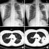
A chest radiograph on admission showed multiple aggregated nodules on her right upper lobe and consolidation with cavity formation on her left upper lobe (Figure 1A). She was diagnosed with M. abscessus pulmonary infection and treated with clarithromycin (1,000 mg/day), cefoxitin (12 g/day), and amikacin (15 mg/kg/day). After admission, she developed a high fever (up to 39℃), which was sustained even during antibiotic treatment, and her chest radiograph showed rapid aggravation (Figure 1B). A chest computed tomography scan also revealed a rapidly aggravating consolidative lesion on the left upper lobe (Figure 1C, D). On the ninth day after admission, moxifloxacin (400 mg/day) was added parenterally. On day 20, imipenem (2,250 mg/day) was substituted for cefoxitin because of pancytopenia (leukocytes 2,700/mm3; hemoglobin 11.1 g/dL; platelets 87,000/mm3) and AST/ALT elevation (AST 146 IU/L, ALT 106 IU/L). On day 25, she underwent a left upper lobectomy because of sustained high fever and a rapidly aggravating consolidative lesion despite high-intensity medical treatment. Immediately after surgery, her fever subsided, AFB stain and culture converted to negativity, and the chest tube was removed 8 days after surgery. She was maintained on four drugs (clarithromycin, amikacin, moxifloxacin, imipenem) for 68 days after surgery. Imipenem, amikacin, and moxifloxacin were sequentially discontinued and she was maintained on clarithromycin alone for an additional 3 months. In total, she was treated with clarithromycin, cefoxitin, amikacin, moxifloxacin, and imipenem for 8.5 months, 19 days, 4 months, 3 months, and 2.5 months, respectively. Drug susceptibility testing (Sensititre; TREK Diagnostic Systems, Cleveland, OH, USA) showed that the clinical isolate was susceptible to linezolid (MIC≤1 µg/mL), clarithromycin (MIC=2 µg/mL), and amikacin (MIC=16 µg/mL), and intermediately susceptible to cefoxitin (MIC=32 µg/mL). The clinical isolate was reidentified by PCR-RFLP and comparative sequence analysis of 16S rDNA and the rpoB and hsp65 genes and finally confirmed to be M. massiliense7. Grossly, the specimen resected from the apicoposterior segment of the left upper lobe appeared as an irregularly shaped firm lesion mixed with a cavitary lesion. Microscopically, chronic granulomatous inflammation with caseous necrosis was observed. There were no visible AFB-positive bacilli and culture of the tissue was negative. She has been followed up for 3 months after completion of treatment without relapse.
Discussion
To the best of our knowledge, this is the first case of M. massiliense pulmonary infection manifesting as acute progressive pneumonia and cured by surgical resection combined with medical treatment. As M. massiliense was only recently separated from the M. abscessus-chelonae group, clinical manifestations of M. massiliense infection are not well characterized. An immunocompetent host with pneumonia caused by M. massiliense, which improved after treatment with clarithromycin and minocycline, has been described1. Our patient, however, showed both rapid progression and unresponsiveness to standard antibiotic treatment. Although 46.5% (59/127) of M. abscessus-chelonae-infected patients in Korea were confirmed with M. massiliense, the clinical characteristics of M. abscessus and M. massiliense pulmonary infections have not been compared. Further studies assessing the clinical characteristics of patients with M. massiliense pulmonary infection are needed.
Paradoxical response or immune reconstitution inflammatory syndrome (IRIS) has been reported not only in TB patients, especially those with acquired immunodeficiency syndrome, but in patients with NTM diseases8,9. In our patient, although a high fever was sustained and a chest radiograph showed rapid aggravation despite antibiotic treatment, a sputum AFB culture immediately before surgery and a culture of the surgical specimen were negative, suggesting that a paradoxical response is more likely than bacteriological unresponsiveness. Hence, immunosuppressive drugs such as corticosteroids may have altered the clinical course and avoided surgical resection. Further studies are needed to clarify this issue.
In summary, we describe here an immunocompetent patient with M. massiliense pulmonary infection presenting as acute progressive pneumonia unresponsive to standard antibiotic treatment, possibly because of a paradoxical response, and improving after surgical resection.
 XML Download