

 PDF
PDF ePub
ePub Citation
 Print
Print


Abstract
Background:
Primary non-Hodgkin's lymphoma of the lung is a rare entity. It is represented commonly as marginal zone B-cell lymphoma of mucosa-associated lymphoid tissue (MALT) type. Although there have been a few reviews of this lymphoma, clinical features, radiologic findings, management and prognosis have not been well defined.
Methods:
We reviewed the medical records of 24 patients with primary pulmonary lymphoma between January 1995 and September 2008; all diagnoses had been confirmed based on pathology.
Results:
The median follow-up time was 42.3 months (range, 0.1∼131.2 months). Five (20.8%) patients were asymptomatic, 17 (70.8%) patients had pulmonary symptoms, and the remaining 2 (8.3%) patients presented with constitutional symptoms. There were 16 (66.7%) patients with MALT lymphoma, 4 (16.7%) patients with diffuse large B-cell lymphoma and 4 (16.7%) patients with lymphoma that had not received a WHO classification. Radiologic findings of primary pulmonary lymphoma were diverse and multiple nodule or consolidation was the most common finding regardless of pathologic lymphoma type. PET scan was carried out in 13 (54.2%) patients and all lesions showed notable FDG uptake. MALT lymphoma showed a trend of better prognosis (3-year survival, 78.8% vs. 70.0%; 5-year survival, 78.8% vs. 52.5%; p=0.310) than non-MALT lymphoma.
Conclusion:
Primary non-Hodgkin's lymphoma of the lung occurs with nonspecific clinical features and radiologic findings. MALT lymphoma is the most common pathologic type of primary pulmonary lymphoma. This entity of lymphoma appears to have a good prognosis and in this study, there was a trend of better outcome than non-MALT lymphoma.
Go to : 
REFERENCES
1.Ferraro P., Trastek VF., Adlakha H., Deschamps C., Allen MS., Pairolero PC. Primary non-Hodgkin's lymphoma of the lung. Ann Thorac Surg. 2000. 69:993–7.

2.Kurtin PJ., Myers JL., Adlakha H., Strickler JG., Lohse C., Pankratz VS, et al. Pathologic and clinical features of primary pulmonary extranodal marginal zone B-cell lymphoma of MALT type. Am J Surg Pathol. 2001. 25:997–1008.
3.Ooi GC., Chim CS., Lie AK., Tsang KW. Computed tomography features of primary pulmonary non-Hodg-kin's lymphoma. Clin Radiol. 1999. 54:438–43.
4.Cordier JF., Chailleux E., Lauque D., Reynaud-Gaubert M., Dietemann-Molard A., Dalphin JC, et al. Primary pulmonary lymphomas: a clinical study of 70 cases in non-immunocompromised patients. Chest. 1993. 103:201–8.
5.Koss MN., Hochholzer L., Nichols PW., Wehunt WD., Lazarus AA. Primary non-Hodgkin's lymphoma and pseudolymphoma of lung: a study of 161 patients. Hum Pathol. 1983. 14:1024–38.
6.Cadranel J., Wislez M., Antoine M. Primary pulmonary lymphoma. Eur Respir J. 2002. 20:750–62.
7.Fiche M., Caprons F., Berger F., Galateau F., Cordier JF., Loire R, et al. Primary pulmonary non-Hodgkin's lymphomas. Histopathology. 1995. 26:529–37.
8.Fisher RI., Dahlberg S., Nathwani BN., Banks PM., Miller TP., Grogan TM. A clinical analysis of two indolent lymphoma entities: mantle cell lymphoma and marginal zone lymphoma (including the mucosa-associated lymphoid tissue and monocytoid B-cell subcategories): a Southwest Oncology Group study. Blood. 1995. 85:1075–82.
9.Habermann TM., Ryu JH., Inwards DJ., Kurtin PJ. Primary pulmonary lymphoma. Semin Oncol. 1999. 26:307–15.
10.Morton LM., Turner JJ., Cerhan JR., Linet MS., Treseler PA., Clarke CA, et al. Proposed classification of lymphoid neoplasms for epidemiologic research from the Pathology Working Group of the International Lymphoma Epidemiology Consortium (InterLymph). Blood. 2007. 110:695–708.
11.Zinzani PL., Tani M., Gabriele A., Poletti V., Stefoni V., Alinari L, et al. Extranodal marginal zone B-cell lymphoma of MALT-type of the lung: single-center experience with 12 patients. Leuk Lymphoma. 2003. 44:821–4.
12.Bégueret H., Vergier B., Parrens M., Lehours P., Laurent F., Vernejoux JM, et al. Primary lung small b-cell lymphoma versus lymphoid hyperplasia: evaluation of diagnostic criteria in 26 cases. Am J Surg Pathol. 2002. 26:76–81.
13.Nicholson AG., Wotherspoon AC., Diss TC., Hansell DM., Du Bois R., Sheppard MN, et al. Reactive pulmonary lymphoid disorders. Histopathology. 1995. 26:405–12.
14.Bienenstock J., Johnston N., Perey DY. Bronchial lymphoid tissue. I. Morphologic characteristics. Lab Invest. 1973. 28:686–92.
15.Kim JH., Lee SH., Park J., Kim HY., Lee SI., Park JO, et al. Primary pulmonary non-Hodgkin's lymphoma. Jpn J Clin Oncol. 2004. 34:510–4.
16.Vanden Eynden F., Fadel E., de Perrot M., de Montpre-ville V., Mussot S., Dartevelle P. Role of surgery in the treatment of primary pulmonary B-cell lymphoma. Ann Thorac Surg. 2007. 83:236–40.
17.Kim JB., Park CK., Park NH., Kum DY., Noh DS., Lee JH, et al. Clinical analysis of primary malignant lymphoma of the lung. Korean J Thorac Cardiovasc Surg. 2007. 40:435–40.
18.Bae YA., Lee KS., Han J., Ko YH., Kim BT., Chung MJ, et al. Marginal zone B-cell lymphoma of bronchus-associated lymphoid tissue: imaging findings in 21 patients. Chest. 2008. 133:433–40.
19.Beal KP., Yeung HW., Yahalom J. FDG-PET scanning for detection and staging of extranodal marginal zone lymphomas of the MALT type: a report of 42 cases. Ann Oncol. 2005. 16:473–80.
20.Hoffmann M., Kletter K., Becherer A., Jäger U., Chott A., Raderer M. 18F-fluorodeoxyglucose positron emission tomography (18F-FDG-PET) for staging and follow-up of marginal zone B-cell lymphoma. Oncology. 2003. 64:336–40.
21.Hoffmann M., Kletter K., Diemling M., Becherer A., Pfeffel F., Petkov V, et al. Positron emission tomography with fluorine-18-2-fluoro-2-deoxy-D-glucose (F18-FDG) does not visualize extranodal B-cell lymphoma of the mucosa-associated lymphoid tissue (MALT)-type. Ann Oncol. 1999. 10:1185–9.
22.Li G., Hansmann ML., Zwingers T., Lennert K. Primary lymphomas of the lung: morphological, immunohistochemical and clinical features. Histopathology. 1990. 16:519–31.
Go to :
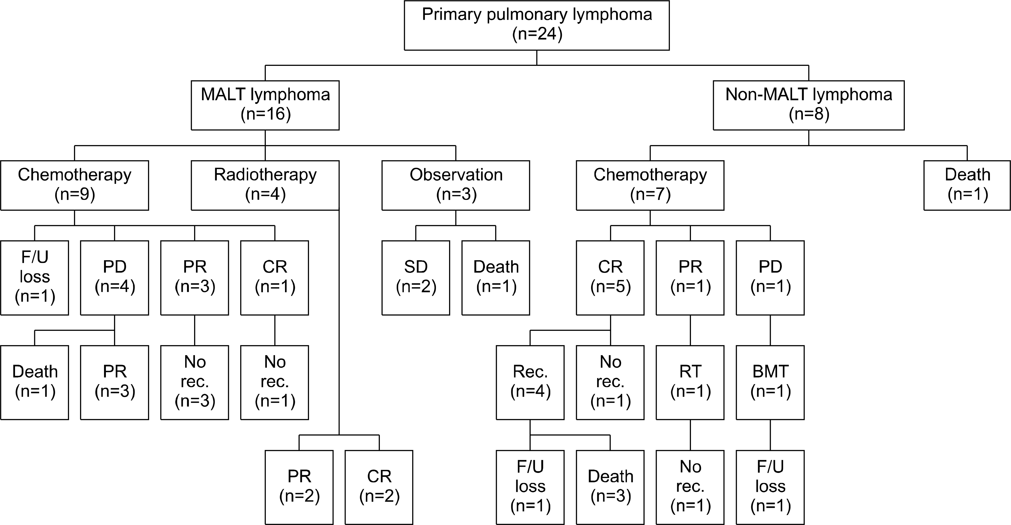 | Figure 1.Clinical courses and final outcomes of the patients with primary pulmonary lymphoma. MALT: mucosa-associated lymphoid tissue; PD: progressive disease; PR: partial response; SD: stable disease; CR: complete remission; Rec.: recurrence; BMT: bone marrow transplantation; RT: radiotherapy; F/U: follow-up. |
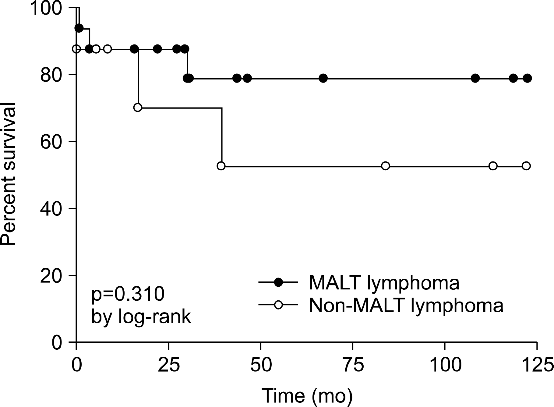 | Figure 2.Comparison of prognosis using Kaplan-Meier survival curves between patients with MALT lymphoma and non-MALT lymphoma. MALT: mucosa-associated lymphoid tissue. |
Table 1.
Characteristics of patients with primary pulmonary lymphoma
Table 2.
Chest CT findings of primary pulmonary lymphoma
Table 3.
Pathologic diagnosis of primary pulmonary lymphoma
| Pathologic diagnosis | No. of patients (%) |
|---|---|
| MALT lymphoma | 16 (66.7) |
| Diffuse large B-cell lymphoma | 4 (16.7) |
| Others∗ | 4 (16.7) |
Table 4.
Comparison of CT findings according to pathologic diagnosis
| MALT lymphoma | Diffuse large B-cell lymphoma | Others∗ | |
|---|---|---|---|
| Multiple nodule or consolidation | 10 (62.5) | 1 (25) | 3 (75) |
| Single nodule or consolidation | 3 (18.8) | 0 | 1 (25) |
| Bronchiectasis and bronchiolitis | 2 (12.5) | 1 (25) | 0 |
| Diffuse interstitial lung disease | 1 (6.3) | 1 (25) | 0 |
| Normal | 0 | 1 (25) | 0 |
| Total | 16 (100) | 4 (100) | 4 (100) |
Table 5.
Comparison of standard uptake value measured by 18F-FDG PET between MALT and non-MALT lymphoma
| Biopsy finding | SUV | p-value |
|---|---|---|
| MALT lymphoma | 3.6±2.5 | 0.334 |
| Non-MALT lymphoma∗ | 8.5±6.2 |
 XML Download