

 PDF
PDF ePub
ePub Citation
 Print
Print


Introduction
Mucus in the airway epithelium plays a pivotal role in defensive mechanisms against airborne chemicals, particles and pathogenic microorganisms. The protective function of airway mucus is due mainly to the viscoelastic property of mucous glycoproteins or mucins. However, any abnormality in the quality or quantity of mucins not only cause altered airway physiology but may also impair host defenses often leading to serious airway pathology as exemplified in chronic bronchitis, cystic fibrosis, asthma, and bronchiectasis1. Therefore, we suggest it is valuable to find the possible activity of controlling (inhibiting) the excess mucin secretion (production) by the components from medicinal plants that have been used for the management of airway diseases. We have tried to investigate the possible activities of some natural products on mucin secretion from cultured airway epithelial cells. As a result of our trial, we previously reported that several natural compounds affected mucin secretion and/or production from airway epithelial cells2,3. According to folk medicine, Glycyrrhiza glabra L. (licorice) has been used for regulating diverse inflammatory diseases including pulmonary diseases4. Carbenoxolone, a steroid-like compound derived from Glycyrrhiza glabra L., was reported to have various biological effects including anti-inflammatory effect5-8. Prunetin, a flavonoid derived from Glycyrrhiza glabra L., showed antioxidant, inhibition of phosphodiesterase, inhibition of aldehyde dehydrogenase and alcohol dehydrogenase activities9-12. Also, according to a number of reports, flavonoids derived from milk thistle, Carduus marianus L., showed hepatoprotective, antioxidant, anti-inflammatory, anti-cancer and immunomodulatory effects13-19. However, to the best of our knowledge, there are no reports about the potential effects of carbenoxolone, prunetin and silibinin on tumor necrosis factor (TNF)-α-induced MUC5AC mucin production and gene expression from human airway epithelial cells. Therefore, in this study, we checked whether carbenoxolone, prunetin and silibinin affect airway mucin production and gene expression stimulated by TNF-α from NCI-H292 cells, a human pulmonary mucoepidermoid cell line.
Materials and Methods
1. Materials
All the chemicals and reagents used in this experiment including carbenoxolone (purity, 98.0%), prunetin (purity, 98.0%) and silibinin (purity, 97.0%) were purchased from Sigma (St. Louis, MO, USA) unless otherwise specified.
2. Cell culture
NCI-H292 cells, a human pulmonary mucoepidermoid carcinoma cell line, were purchased from the American Type Culture Collection (ATCC, Manassas, VA, USA) and cultured in RPMI 1640 supplemented with 10% fetal bovine serum (FBS) in the presence of penicillin (100 units/mL), streptomycin (100 µg/mL) and HEPES (25 mM) at 37℃ in a humidified, 5% CO2/95% air, water-jacketed incubator. For serum deprivation, confluent cells were washed twice with phosphate-buffered saline (PBS) and recultured in RPMI 1640 with 0.2% fetal bovine serum for 24 hours.
3. Treatment of cells with carbenoxolone, prunetin, and silibinin
After 24 hours of serum deprivation, cells were pretreated with carbenoxolone (1, 10, 100 µM), prunetin (1, 10, 100 µM), and silibinin (1, 10, 100 µM) for 30 minutes and treated with TNF-α (0.2 nM) for 24 hours in serum-free RPMI 1640. After 24 hours, cells were lysed with buffer solution containing 20 mM Tris, 0.5% NP-40, 250 mM NaCl, 3 mM EDTA, 3 mM EGTA and protease inhibitor cocktail (Roche Diagnostics, Indianapolis, IN, USA) and collected to measure the production of MUC5AC protein (in 24-well culture plate). The total RNA was extracted for measuring the expression of MUC5AC gene (in 6-well culture plate) by using reverse transcription-polymerase chain reaction (RT-PCR).
4. MUC5AC mucin analysis using ELISA
MUC5AC protein was measured by using enzyme-linked immunosorbent assay (ELISA). Cell lysates were prepared with PBS at 1 : 10 dilution, and 100 µL of each sample was incubated at 42℃ in a 96-well plate, until dry. Plates were washed three times with PBS and blocked with 2% BSA (fraction V) for 1 hour at room temperature. Plates were again washed three times with PBS and then incubated with 100 µL of 45M1, a mouse monoclonal MUC5AC antibody (NeoMarkers, Freemont, CA, USA) (1 : 200), which was diluted with PBS containing 0.05% Tween 20 and dispensed into each well. After 1 hour, the wells were washed three times with PBS, and 100 µL of horseradish peroxidase-goat anti-mouse IgG conjugate (1 : 3,000) was dispensed into each well. After 1 hour, plates were washed three times with PBS. Color reaction was developed with 3,3',5,5'-tetramethylbenzidine (TMB) peroxide solution and stopped with 1 N H2SO4. Absorbance was read at 450 nm.
5. Total RNA isolation and RT-PCR
Total RNA was isolated by using Easy-BLUE Extraction Kit (iNtRON Biotechnology Inc., Seongnam, Korea) and reverse transcribed by using AccuPower RT Premix (BIONEER Co., Daejeon, Korea) according to the manufacturer's instructions. 2 µg of total RNA was primed with 1 µg of oligo (dT) in a final volume of 50 µL (RT reaction). 2 µL of RT reaction product was PCR amplified in a 25 µL by using Thermorprime Plus DNA Polymerase (ABgene, Rochester, NY, USA). Primers for MUC5AC were (forward) 5'-TGA TCA TCC AGC AGG GCT-3' and (reverse) 5'-CCG AGC TCA GAG GAC ATA TGG G-3'. The size of expected fragment amplified by PCR was 458 bp. As quantitative controls, primers for Rig/S15 rRNA, which encodes a small ribosomal subunit protein, a housekeeping gene that was constitutively expressed, were used. Primers for Rig/S15 were (forward) 5'-TTC CGC AAG TTC ACC TAC C-3' and (reverse) 5'-CGG GCC GGC CAT GCT TTA CG-3'. The size of expected fragment amplified by PCR was 361 bp. The PCR mixture was denatured at 94℃ for 2 minutes followed by 40 cycles at 94℃ for 30 seconds, 60℃ for 30 seconds and 72℃ for 45 seconds. After PCR, 5 µL of PCR products were subjected to 1% agarose gel electrophoresis and visualized with ethidium bromide under a transilluminator.
Results
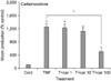
1. Effect of carbenoxolone on TNF-α-induced MUC5AC mucin production from NCI-H292 cells
As can be seen in Figure 1, carbenoxolone inhibited TNF-α-induced MUC5AC mucin production. The amounts of MUC5AC mucin in the cells of carbenoxolone-treated cultures were 100±10%, 1,250±98%, 1,235±108%, 1,124±76%, and 500±48% for control, TNF-α 0.2 nM only, carbenoxolone 10-6 M+TNF-α, carbenoxolone 10-5 M+TNF-α and carbenoxolone 10-4 M+TNF-α, respectively (Figure 1).
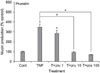
2. Effect of prunetin on TNF-α-induced MUC5AC mucin production from NCI-H292 cells
As can be seen in Figure 2, prunetin also inhibited TNF-α-induced MUC5AC mucin production, dose-dependently. The amounts of MUC5AC mucin in the cells of prunetin-treated cultures were 100±6%, 349±29%, 284±30%, 92±9%, and 69±5% for control, TNF-α 0.2 nM only, prunetin 10-6 M+TNF-α, prunetin 10-5 M+TNF-α and prunetin 10-4 M+TNF-α, respectively (Figure 2).
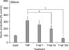
3. Effect of silibinin on TNF-α-induced MUC5AC mucin production from NCI-H292 cells
As can be seen in Figure 3, silibinin inhibited TNF-α-induced MUC5AC mucin production, dose-dependently. The amounts of MUC5AC mucin in the cells of silibinin-treated cultures were 100±7%, 317±29%, 257±30%, 198±13%, and 59±5% for control, TNF-α 0.2 nM only, silibinin 10-6 M+TNF-α, silibinin 10-5 M+TNF-α and silibinin 10-4 M+TNF-α, respectively (Figure 3).
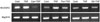
4. Effects of carbenoxolone, prunetin and silibininon TNF-α-induced MUC5AC gene expression from NCI-H292 cells
As can be seen in Figure 4, carbenoxolone, prunetin and silibinin inhibited TNF-α-induced MUC5AC gene expression at the concentration of 10-4 M, respectively.
Discussion
MUC5AC was reported to be mainly expressed in goblet cells existing in the airway surface epithelium among the twenty genes coding human mucins (MUC)20. Also, TNF-α is a well-known stimulator of secretion and gene expression of airway mucin21-23. TNF-α level in sputum was reported to be increased, with further increases during exacerbation of diseases24,25. TNF-α converting enzyme (TACE) mediated MUC5AC mucin expression in cultured human airway epithelial cells22 and TNF-α induced MUC5AC gene expression in normal human airway epithelial cells23. It also induced mucin secretion from guinea pig tracheal epithelial cells21. On the basis of these reports, in this study, we tried to test the possible effects of carbenoxolone, prunetin and silibinin on TNF-α-induced MUC5AC mucin production and gene expression from NCI-H292 cells, a human pulmonary mucoepidermoid cell line, which are frequently used for the purpose of elucidating intracellular signaling pathways involved in airway mucin production and gene expression22,26. As can be seen in results, carbenoxolone, prunetin and silibinin inhibited the production of MUC5AC mucin protein induced by TNF-α. In the result of experiment shown in Figure 1, TNF-α induced MUC5AC mucin production more potently than mucin productions in the other two results of experiments shown in Figures 2 and 3. Actually, based on the preliminary experiments performed by our group, the efficacy of stimulation of mucin production by TNF-α varied depending on the experimental conditions, although the exact cause of this phenomenon is unclear at present. On the other hand, the three compounds inhibited the expression of MUC5AC mucin gene induced by TNF-α. This result suggests that carbenoxolone, prunetin and silibinin can regulate mucin gene expression and production of mucin protein, by directly acting on airway epithelial cells. The underlying mechanisms of action of these three compounds on MUC5AC production and gene expression are not clear at present, although we are trying to investigate whether these three compounds act as regulators of NF-kB signaling pathway activated by TNF-α in mucin-producing NCI-H292 cells. Taken together, the inhibitory actions of carbenoxolone, prunetin and silibinin on airway mucin production and gene expression might explain, at least in part, the traditional use of G. glabra L. as anti-inflammatory agent and mucoregulator for airway inflammatory diseases, in oriental medicine and the folk use of Carduus marianus L. as anti-inflammatory agent. We suggest it is valuable to find the natural products that have specific inhibitory effects on mucin production and/or gene expression - in view of both basic and clinical sciences - and the result from this study suggest a possibility of using carbenoxolone, prunetin and silibinin as new efficacious mucoregulators for respiratory diseases, although further studies are essential.
 XML Download