

 PDF
PDF ePub
ePub Citation
 Print
Print


Abstract
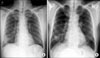
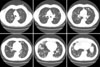
Novel influenza A (H1N1) virus is a common pathogen of febrile respiratory infection recently. Here, we report the case of a 63-year-old male patient who presented with 3 days' ongoing cough and fever. He was diagnosed with novel influenza A (H1N1) pneumonia by real-time reverse-transcriptase-polymerase-chain-reaction (rRT-PCR). During treatment for novel influenza A (H1N1), his symptoms and radiologic findings improved initially, but multiple lung nodules developed subsequently and found on chest x-ray (on the 5th hospital day). Mycobacterium abscessus was isolated repeatedly from sputum and bronchoalveolar lavage fluid. To our knowledge, this is the first reported case of Mycobacterium abscessus lung disease in a patient with H1N1 influenza pneumonia.
Figures and Tables
References
1. Influenza-like illness in the United States and Mexico [Internet]. World Health Organization. c2010. cited 2009 Apr 24. Geneva: World Health Organization;Available from:
http://www.who.int/csr/don/2009_04_24/en.
2. Pandemic (H1N1) 2009 - update 83 [Internet]. World Health Organization. c2010. cited 2010 Jan 15. Geneva: World Organization;Available from:
http://www.who.int/csr/don/2010_01_15/en.
3. Rothberg MB, Haessler SD. Complications of seasonal and pandemic influenza. Crit Care Med. 2009. 38:Suppl 4. e91–e97.
4. Kantzler GB, Lauteria SF, Cusumano CL, Lee JD, Ganguly R, Waldman RH. Immunosuppression during influenza virus infection. Infect Immun. 1974. 10:996–1002.
5. Wallace RJ Jr. Recent changes in taxonomy and disease manifestations of the rapidly growing mycobacteria. Eur J Clin Microbiol Infect Dis. 1994. 13:953–960.
6. Wallace RJ Jr, Swenson JM, Silcox VA, Good RC, Tschen JA, Stone MS. Spectrum of disease due to rapidly growing mycobacteria. Rev Infect Dis. 1983. 5:657–679.
7. Griffith DE, Girard WM, Wallace RJ Jr. Clinical features of pulmonary disease caused by rapidly growing mycobacteria. An analysis of 154 patients. Am Rev Respir Dis. 1993. 147:1271–1278.
8. Ingram CW, Tanner DC, Durack DT, Kernodle GW Jr, Corey GR. Disseminated infection with rapidly growing mycobacteria. Clin Infect Dis. 1993. 16:463–471.
9. Koh WJ, Kwon OJ, Ham HS, Suh GY, Chung MP, Kim HJ, et al. Clinical significance of nontuberculous mycobacteria isolated from respiratory specimens. Korean J Med. 2003. 65:10–21.
10. Collins CH, Grange JM, Yates MD. Mycobacteria in water. J Appl Bacteriol. 1984. 57:193–211.
11. Wolinsky E, Rynearson TK. Mycobacteria in soil and their relation to disease-associated strains. Am Rev Respir Dis. 1968. 97:1032–1037.
12. Griffith DE, Aksamit T, Brown-Elliott BA, Catanzaro A, Daley C, Gordin F, et al. An official ATS/IDSA statement: diagnosis, treatment, and prevention of nontuberculous mycobacterial diseases. Am J Respir Crit Care Med. 2007. 175:367–416.
13. Koh WJ, Kwon OJ. Diagnosis and treatment of nontuberculous mycobacterial lung disease. Korean J Med. 2008. 74:120–131.
14. Jeon K, Kwon OJ, Lee NY, Kim BJ, Kook YH, Lee SH, et al. Antibiotic treatment of Mycobacterium abscessus lung disease: a retrospective analysis of 65 patients. Am J Respir Crit Care Med. 2009. 180:896–902.
15. Daley CL, Griffith DE. Pulmonary disease caused by rapidly growing mycobacteria. Clin Chest Med. 2002. 23:623–632. vii
 XML Download