

 PDF
PDF ePub
ePub Citation
 Print
Print


Introduction
Prostatic cancer is the most common cancer of men in western countries1 while its incidence and mortality in Asia were estimated to be quite lower although there has been a recent trend towards increasing incidence of prostate cancer in that area2. Bones such as pelvis, femur, spines and ribs are frequently involved by hematogenous metastasis through venous plexus of Batson3. Pulmonary metastasis is also not uncommon4.
Symptoms from prostatic cancer are rarely present at the time of diagnosis in most patients. Therefore, it was often detected due to problems of metastatic site5. In addition, because presenting features of prostatic cancer are sometimes very unusual, there is the risk of wrong diagnosis. Here, we report a patient with prostatic cancer presenting as isolated large lung mass. Because initially, there were no diagnostic clues suggesting the primary site, it almost mimicked the primary lung cancer.
Case Report
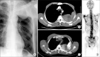
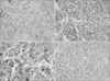
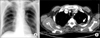
A large left upper lung mass was incidentally found on chest radiograph in a 59-year-old man with no history of smoking (Figure 1A). He was previously healthy and did not present any specific symptoms related with respiratory, gastro-intestinal, skeletal and genitor-urinary systems. A 9.4 cm-sized mass along posterior pleural surface in the left upper lobe without any enlarged mediastinal lymph node was found on chest computed tomography (CT) (Figure 1B). 18F-FDG PET revealed hypermetabolic lesions in lung, abdominal lymph nodes and multiple bones (Figure 1C). Extensive bone metastasis was also confirmed by a bone scan (Figure 1D). However, there was no abnormal uptake in other organs including the gastro-intestinal and genitor-urinary tracts. A sample of the lung mass was obtained by percutaneous needle aspiration biopsy. Pathologic examination revealed adenocarcinoma (Figure 2A). Although the diagnosis of lung cancer with multiple metastases was probable, we decided to check tumor markers to rule out lung metastasis from other primary cancers because the immunostaining for thyroid transcription factor-1 (TTF-1) was negative (Figure 2B). Unexpectedly, the serum PSA level was high (5148.0 ng/mL; normal range, 0~4 ng/mL), raising the possibility of prostatic cancer. A prostatic mass was detected by ultrasound and a trans-rectal biopsy was done. Pathologic examination of the specimen showed adenocarcinoma with a Gleason score 4+5, which involved 11 of the 12 cores taken from the prostate (Figure 2C). Lung and prostate tissues displayed the same morphology and were both positive for PSA immmunostaining, leading to the final diagnosis of prostate cancer with multiple metastases (Figure 2D). Therapy with luteinizing hormone-releasing hormone agonist (3.6 mg, once monthly, subcutaneous injection) was started. A follow-up examination with chest radiography and CT after 1 month showed that the lung lesion had markedly decreased in size (Figure 3). Serum PSA level had also decreased to 143.9 ng/mL.
Discussion
This report clearly showed that physicians should be very cautious before making a diagnosis of lung cancer because of rare presentation as a lung mass in other site malignancy. Hence, it is required to carefully review all data before final diagnosis for discovery of something unusual in lung cancer, especially, with adenocarcinoma histology because the primary adenocarcinoma can be more silently present inside body. Adenocarcinoma has become the most common histology of lung cancer accounting for more than 30%6,7. It is expected that the proportion of adenocarcinoma would continue to increase. Moreover, recent use of low-dose spiral CT for lung cancer screening would detect more metastasis of other hidden primary cancer to lung including early, isolated lesion in addition to early lung cancer8,9. Therefore, there would be more chances for physicians to encounter patients like our case reported here.
We performed TTF-1 immunostaining because we thought it is not common for lung cancer to metastasize to multiple bones and abdominal lymph nodes without mediastinal involvement. In addition, the differentiation from malignant pleural mesothelioma was also needed because the tumor mass broadly contacted with pleura surface, and because malignant pleural mesothelioma and peripheral adenocarcinoma of the lung often show similar clinical and radiological characteristics and even similar microscopic findings 10,11. TTF-1 immunostaining is a very sensitive and specific method in the differential diagnosis of primary and metastatic lung adenocarcinoma12 and also can be useful to exclude mesothelioma13. It helped us in the search for another primary site of cancer.
Should TTF-1 immunostaining be routinely done before diagnosis of lung adenocarcinoma? It is a quite controversial question although the procedure can be done easily and without substantial cost and it would help to lessen the already-low risk of embarking on the wrong treatment. More accumulation of clinical experiences about hidden adenocarcinoma presenting a lung mass seems to be able to answer it.
 XML Download