

 PDF
PDF ePub
ePub Citation
 Print
Print


Abstract
Pulmonary arteriovenous malformation (PAVM) is a rare pulmonary vascular anomaly due to an abnormal communication between the pulmonary artery and vein. The most common presenting symptom is a dyspnea on exertion related to this right-to-left shunt. If left untreated, PAVM has been known to result in serious complications. Incomplete pulmonary capillary network can be the cause of cerebral abscesses and other non-infectious neurological complications, such as stroke and transient ischemic attacks due to paradoxic embolism Transcatheter embolotherapy, using coils or balloons, has replaced surgical resection as the treatment of choice for PAVM. However, the risk of device embolization has limited the use of coil embolotherapy, while the size of PAVM is huge. Recently, Amplatzer® Vascular Plug has been proposed as an alternative endovascular occlusion device for arteriovenous malformation. We report a case of 81-year-old male patient with a giant PAVM, which was successfully treated by transcatheter embolotherapy using the Amplatzer® Vascular Plug.
Figures and Tables
Figure 1
(A) Chest radiograph on admission showed a circumscribed mass shadow in the left lower hemithorax. (B~D) Computed tomography (CT) scan of chest revealed centrilobular emphysema on whole lung fields and a large saccular dilatation of left pulmonary artery directly draining to left pulmonary vein in the left lower lobe.
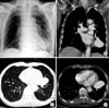
Figure 2
(A) Left pulmonary arteriogram showed large pulmonary arteriovenous malformation. (B, C) A 12 mm Amplatzer® Vascular Plug was inserted and deployed via 7F guiding catheter. (D) Pulmonary arteriovenous malformation was not seen on follow-up arteriogram.
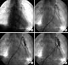
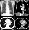
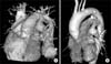
Figure 3
The size of previously described mass shadow of pulmonary arteriovenous malformation in the left lower lobe decreased on chest radiograph (A) and chest CT (B~D) after transcatheter embolotherapy with 12 mm Amplatzer® Vascular Plug located at feeding artery of pulmonary arteriovenous malformation.
References
1. Gossage JR, Kanj G. Pulmonary arteriovenous malformations: a state of the art review. Am J Respir Crit Care Med. 1998. 158:643–661.
2. Loke GP, Story DA, Liskaser F, Seevanayagam S. Pulmonary arteriovenous malformation causing massive haemoptysis and complicated by coronary air embolism. Anaesth Intensive Care. 2006. 34:75–78.
3. Khurshid I, Downie GH. Pulmonary arteriovenous malformation. Postgrad Med J. 2002. 78:191–197.
4. Pierucci P, Murphy J, Henderson KJ, Chyun DA, White RI Jr. New definition and natural history of patients with diffuse pulmonary arteriovenous malformations: twenty-seven-year experience. Chest. 2008. 133:653–661.
5. Jung JY, Lim JK, Chun SW, Suh WN, Kim DJ, Lee KH, et al. A case of video-assisted thoracoscopic pneumonectomy for unilateral diffuse pulmonary arteriovenous malformation. Tuberc Respir Dis. 2006. 61:585–590.
6. Kim MD, Kim JS, Lim CY. Treatment of pulmonary arteriovenous malformation using platinum coils: case report. J Korean Radiol Soc. 2005. 52:113–116.
7. Han YM, Song HY, Lee JM, Chung JY, Lee SY, Chung GH, et al. Pulmonary arteriovenous malformation treatment with detachable balloon: a case report. J Korean Radiol Soc. 1996. 34:595–598.
8. Luthra S, Antippa P, Tatoulis J. Pulmonary arteriovenous aneurysm as manifestation of Osler-Weber-Rendu syndrome. Heart Lung Circ. 2008. 17:336–339.
9. Gezer S, Turut H, Oz G, Demirag F, Tastepe I. Acquired pulmonary arteriovenous malformation secondary to hydatid cyst operation. Thorac Cardiovasc Surg. 2007. 55:462–463.
10. Kim CH, Kim TH, Park JB, Jeong JP, Ko YC, Lee SU, et al. A case of diffuse pulmonary arteriovenous malformation with multiple nodules on chest X-ray. Chonnam Med J. 2005. 41:233–236.
11. Ones T, Dede F, Erdim R, Erdil TY, Inanir S, Yuksel M, et al. Quantitative shunt imaging in the evaluation of therapeutic surgery in a patient with pulmonary arteriovenous malformation. Ann Thorac Surg. 2008. 86:649–651.
12. Cil B, Canyigit M, Ozkan OS, Pamuk GA, Dogan R. Bilateral multiple pulmonary arteriovenous malformations: endovascular treatment with the Amplatzer Vascular Plug. J Vasc Interv Radiol. 2006. 17:141–145.
13. Hinterseer M, Becker A, Barth AS, Kozlik-Feldmann R, Wintersperger BJ, Behr J. Interventional embolization of a giant pulmonary arteriovenous malformation with right-left-shunt associated with hereditary hemorrhagic telangiectasia. Clin Res Cardiol. 2006. 95:174–178.
14. Abdel Aal AK, Hamed MF, Biosca RF, Saddekni S, Raghuram K. Occlusion time for Amplatzer vascular plug in the management of pulmonary arteriovenous malformations. AJR Am J Roentgenol. 2009. 192:793–799.
15. Kim JH, Park OS, Lee KW, Yun SH, Kang DG, Ko YC, et al. A case of embolotherapy of diffuse pulmonary arteriovenous malformation using Amplatzer Vascular Plugs. Chonnam Med J. 2006. 42:144–147.
 XML Download