

 PDF
PDF ePub
ePub Citation
 Print
Print


Abstract
Background
Micro computed tomography (CT) is rapidly developing as an imaging tool, especially for mice, which have become the experimental animal of choice for many pulmonary disease studies. We evaluated the usefulness of micro CT for evaluating lung fibrosis in the murine model of bleomycin-induced lung inflammation and fibrosis.
Methods
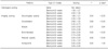
The control mice (n=10) were treated with saline. The murine model of lung fibrosis (n=60) was established by administering bleomycin intra-tracheally. Among the 70 mice, only 20 mice had successful imaging analyses. We analyzed the micro CT and pathological findings and examined the correlation between imaging scoring in micro CT and histological scoring of pulmonary inflammation or fibrosis.
Results
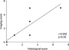
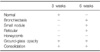
The control group showed normal findings on micro CT. The abnormal findings on micro CT performed at 3 weeks after the administration of bleomycin were ground-glass opacity (GGO) and consolidation. At 6 weeks after bleomycin administration, micro CT showed various patterns such as GGO, consolidation, bronchiectasis, small nodules, and reticular opacity. GGO (r=0.84) and consolidation (r=0.69) on micro CT were significantly correlated with histological scoring that reflected pulmonary inflammation (p<0.05). In addition, bronchiectasis (r=0.63) and reticular opacity (r=0.83) on micro CT shown at 6 weeks after bleomycin administration correlated with histological scoring that reflected lung fibrosis (p<0.05).
Figures and Tables
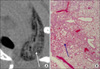
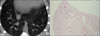 | Figure 1Micro CT finding (A) and pathologic finding (B) of saline-treated mice. Both micro CT scan and lung tissue show normal findings (H&E stain, ×25).
|
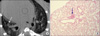 | Figure 2Micro CT finding (A) and pathologic finding (B) of bleomycin-treated mice that is classified as small nodule according to the pattern of micro CT finding. Micro CT scanning and histological sampling were performed at 6 weeks after the administration of bleomycin solution. (A) Micro CT shows two small nodules (white arrow) in right lower lobe on micro CT. (B) Histological finding shows an aggregation of inflammatory cells (black arrow) with fibroblast around bronchus (H&E stain, ×25).
|
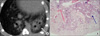 | Figure 3Micro CT finding (A) and pathologic finding (B) of bleomycin-treated mice that is classified as consolidation according to the pattern of micro CT finding. Micro CT scanning and histological sampling were performed at 3 weeks after the administration of bleomycin solution. (A) Micro CT shows the consolidation (white arrow) in left lower lobe. (B) Histological finding shows many inflammatory cells around bronchi, alveoli (black arrow), and alveolar septa (H&E stain, ×25).
|
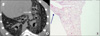 | Figure 4Micro CT finding (A) and pathologic finding (B) of bleomycin-treated mice that show many patterns related with lung fibrosis on micro CT. Micro CT scanning and histological sampling were performed at 6 weeks after the administration of bleomycin solution. (A) Micro CT shows diffuse ground-glass opacity, reticular opacity (white arrow), bronchiolectasis, and small nodule in left lower lobe. (B) Histological finding shows dense collagen deposition and interstitial fibrosis (black arrow) (H&E stain, ×25).
|
 | Figure 5Micro CT finding (A) and pathologic finding (B) of bleomycin-treated mice that have severe lung fibrosis. Micro CT scanning and histological sampling were performed at 6 weeks after the administration of bleomycin solution. (A) Micro CT shows diffuse ground-glass opacity with reticular pattern (white arrow) in left lower lobe. (B) Histological finding shows a reticular pattern (black arrow) due to severe interstitial fibrosis with destruction (H&E stain, ×25).
|
References
1. Gross TJ, Hunninghake GW. Idiopathic pulmonary fibrosis. N Engl J Med. 2001. 345:517–525.
2. Moeller A, Ask K, Warburton D, Gauldie J, Kolb M. The bleomycin animal model: a useful tool to investigate treatment options for idiopathic pulmonary fibrosis? Int J Biochem Cell Biol. 2008. 40:362–382.
3. Sleijfer S. Bleomycin-induced pneumonitis. Chest. 2001. 120:617–624.
4. Ritman EL. Micro-computed tomography of the lungs and pulmonary-vascular system. Proc Am Thorac Soc. 2005. 2:477–480. 501
5. Ritman EL. Micro-computed tomography-current status and developments. Annu Rev Biomed Eng. 2004. 6:185–208.
6. Johnson KA. Imaging techniques for small animal imaging models of pulmonary disease: micro-CT. Toxicol Pathol. 2007. 35:59–64.
7. Lee HJ, Goo JM, Kim NR, Kim MA, Chung DH, Son KR, et al. Semiquantitative measurement of murine bleomycin-induced lung fibrosis in in vivo and postmortem conditions using microcomputed tomography: correlation with pathologic scores--initial results. Invest Radiol. 2008. 43:453–460.
8. Cavanaugh D, Travis EL, Price RE, Gladish G, White RA, Wang M, et al. Quantification of bleomycin-induced murine lung damage in vivo with micro-computed tomography. Acad Radiol. 2006. 13:1505–1512.
9. Genovese T, Cuzzocrea S, Di Paola R, Mazzon E, Mastruzzo C, Catalano P, et al. Effect of rosiglitazone and 15-deoxy-Delta12, 14-prostaglandin J2 on bleomycin-induced lung injury. Eur Respir J. 2005. 25:225–234.
10. Ashcroft T, Simpson JM, Timbrell V. Simple method of estimating severity of pulmonary fibrosis on a numerical scale. J Clin Pathol. 1988. 41:467–470.
11. Langheinrich AC, Leithäuser B, Greschus S, Von Gerlach S, Breithecker A, Matthias FR, et al. Acute rat lung injury: feasibility of assessment with micro-CT. Radiology. 2004. 233:165–171.
12. Ford NL, Wheatley AR, Holdsworth DW, Drangova M. Optimization of a retrospective technique for respiratory-gated high speed micro-CT of free-breathing rodents. Phys Med Biol. 2007. 52:5749–5769.
 XML Download