Abstract
Background:
A diagnosis and treatment of chest wall tuberculosis (CWTB) is both difficult and controversial. The aim of this study was to collect information on the optimal treatment for CWTB.
Methods:
The clinical features, radiographic findings, and treatment outcomes of 26 patients, who underwent surgery and were diagnosed histopathologically, were retrospectively analyzed.
Results:
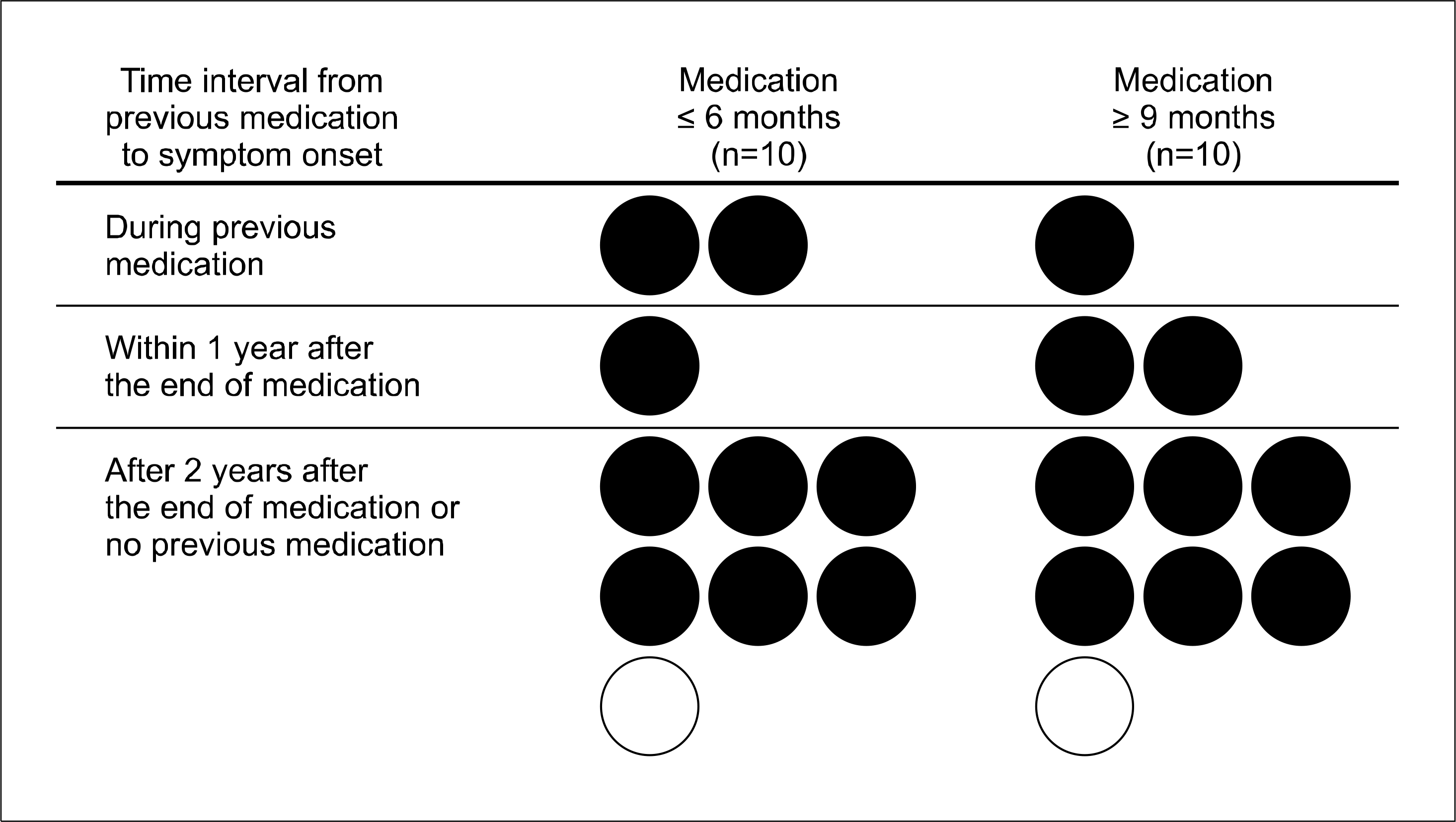
The most common presenting symptom was a palpable mass found in 24 patients (92.3%). In all patients, CT revealed a soft tissue mass that was accompanied by a central low density, with or without peripheral rim enhancement. The sensitivity and specificity of the bone scintigram for bone involvement were 87.5% and 100%, respectively. CWTB was diagnosed preoperatively by aspiration cytology and smear for acid‐fast bacilli in five out of 11 patients. Twenty‐three patients (88.5%) underwent a radical excision and three underwent incision/drainage or an incisional biopsy. The duration of antituberculous medication was 7.5±3.98 months with a follow‐up period of 28.2±26.74 months. Among the 20 patients who completed their treatment, nine received chemotherapy for six months or less and 11 received chemotherapy for nine months or more. Two patients had a recurrence four and seven months after starting their medication.
Go to : 
REFERENCES
1.Faure E., Souilamas R., Riquet M., Chehab A., Le Pimpec-Barthes F., Manac'h D, et al. Cold abscess of the chest wall: a surgical entity? Ann Thorac Surg. 1998. 66:1174–8.

2.Morris BS., Maheshwari M., Chalwa A. Chest wall tuberculosis: a review of CT appearances. Br J Radiol. 2004. 77:449–57.

3.Hulnick DH., Naidich DP., McCauley DI. Pleural tuberculosis evaluated by computed tomography. Radiology. 1983. 149:759–65.

5.Mathlouthi A., Ben M'Rad S., Merai S., Friaa T., Mestiri I., Ben Miled K, et al. Tuberculosis of the thoracic wall: presentation of 4 personal cases and review of the literature. Rev Pneumol Clin. 1998. 54:182–6.
6.Eid A., Chaudry N., el-Ghoroury M., Hawasli A., Salot WL., Khatib R. Multifocal musculoskeletal cystic tuberculosis without systemic manifestations. Scand J Infect Dis. 1994. 26:761–4.

7.Garcia S., Combalia A., Serra A., Segur JM., Ramon R. Unusual locations of osteoarticular tuberculosis. Arch Orthop Trauma Surg. 1997. 116:321–3.

8.Chang DS., Rafii M., McGuinness G., Jagirdar JS. Primary multifocal tuberculous osteomyelitis with involvement of the ribs. Skeletal Radiol. 1998. 27:641–5.

9.Martini M., Ouahes M. Bone and joint tuberculosis: a review of 652 cases. Orthopedics. 1988. 11:861–6.

10.Tatelman M., Drouillard EJ. Tuberculosis of the ribs. Am J Roentgenol Radium Ther Nucl Med. 1953. 70:923–35.
11.Paik HC., Chung KY., Kang JH., Maeng DH. Surgical treatment of tuberculous cold abscess of the chest wall. Yonsei Med J. 2002. 43:309–14.

12.Hsu HS., Wang LS., Wu YC., Fahn HJ., Huang MH. Management of primary chest wall tuberculosis. Scand J Thorac Cardiovasc Surg. 1995. 29:119–23.

13.Khalil A., Le Breton C., Tassart M., Korzec J., Bigot J., Carette M. Utility of CT scan for the diagnosis of chest wall tuberculosis. Eur Radiol. 1999. 9:1638–42.

14.Ward AS. Superficial abscess formation: an unusual presenting feature of tuberculosis. Br J Surg. 1971. 58:540–3.

15.Chen CH., Shih JF., Wang LS., Perng RP. Tuberculous subcutaneous abscess: an analysis of seven cases. Tuber Lung Dis. 1996. 77:184–7.

16.Blunt SB., Harries MG. Discrete pleural masses without effusion in a young man: an unusual presentation of tuberculosis. Thorax. 1989. 44:436–7.

17.Chang JH., Kim SK., Lee WY. Diagnostic issues in tuberculosis of the ribs with a review of 12 surgically proven cases. Respirology. 1999. 4:249–53.

18.Dutt AK., Moers D., Stead WW. Short-course chemotherapy for extrapulmonary tuberculosis: nine years' experience. Ann Intern Med. 1986. 104:7–12.
19.Brown RB., Trenton J. Chronic abscesses and sinuses of the chest wall: the treatment of costal chondritis and sternal osteomyelitis. Ann Surg. 1952. 135:44–51.
Go to : 
 | Figure 1.Chest CT scan showing a soft tissue mass (arrow) with a central low density and with peripheral rim enhancement in the inner and outer sides of the ribs (A), and only in the outer side (B). The subcuatenous fat layer (arrow) was only involved in one lesion (C). |
 | Figure 2.Six- or less than six-month regimen versus nine- or more than nine-month regimen, which was stratified by time intervals from the end of the previous medication to symptom onset of chest wall tuberculosis. The numbers mean the duration of antituberculous medication, and solid (●) and blank (○) circles indicate cured and recurred cases, respectively. |
Table 1.
Clinical characteristics of the patients (n=26)
Table 2.
Chest CT findings
Table 3.
Bone scintigram (n=10)
| Surgical or pathological bone involvement | Bone scintigram | |
|---|---|---|
| Positive hot uptake | Negative hot uptake | |
| Present | 7 | 1 |
| Absent | 0 | 2 |
Table 4.
Microbiologic and cytopathologic results


 PDF
PDF ePub
ePub Citation
 Print
Print


 XML Download