

 PDF
PDF ePub
ePub Citation
 Print
Print


Abstract
The stomach is a rare site for metastasis, with autopsy incidence rates of 0.2% to 1.7%. This low rate makes diagnosis of metastatic gastric cancer challenging for clinicians. The authors report a case of a 64-year-old man diagnosed with gastric metastasis of primary lung adenocarcinoma that was initially mistaken for primary gastric cancer, as well as a review of the medical literature.
Figures and Tables
Figure 1
Initial gastrofiberscopy showed 3.2 cm sized polypoid mass with ulceration in the body of stomach.
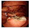
Figure 2
(A) Chest X-ray taken during the admission for gastrectomy showed a ground-glass opacity (GGO) lesion in the right upper lobe. (B) CT scan taken 2 months after subtotal gastrectomy showed a GGO lesion with solid portion in the right upper lobe. (C, D) Chest X-ray and CT scan taken 11 months after right upper lobectomy showed a mass with surrounding ground-glass opacity in the left lower lobe.
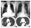
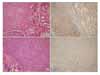
Figure 3
(A) Hematoxylin and eosin (H&E) stain in the lung tissue obtained by percutaneous needle biopsy (×100). A papillary pattern and undifferentiated diffuse proliferation, suggesting poorly differentiated adenocarcinoma are observed. (B) Immunohistochemical stain for thyroid transcription factor-1 (TTF-1) in the lung tissue obtained by percutaneous needle biopsy (×100). The strong positive staining with TTF-1 is observed. (C) H&E stain in the gastrectomy specimen (×100). The tumor cells of stomach are similar to the adenocarcinoma observed in the lung tissue. (D) Immunohistochemical stain for TTF-1 in the gastrectomy specimen (×100). The strong positive staining with TTF-1 is observed.
References
1. Kadakia SC, Parker A, Canales L. Metastatic tumors to the upper gastrointestinal tract: endoscopic experience. Am J Gastroenterol. 1992. 87:1418–1423.
2. Menuck LS, Amberg JR. Metastatic disease involving the stomach. Am J Dig Dis. 1975. 20:903–913.
3. Wu MH, Lin MT, Lee PH. Clinicopathological study of gastric metastases. World J Surg. 2007. 31:132–136.
4. Green LK. Hematogenous metastases to the stomach: a review of 67 cases. Cancer. 1990. 65:1596–1600.
5. Oda , Kondo H, Yamao T, Saito D, Ono H, Gotoda T, et al. Metastatic tumors to the stomach: analysis of 54 patients diagnosed at endoscopy and 347 autopsy cases. Endoscopy. 2001. 33:507–510.
6. Hsu CC, Chen JJ, Changchien CS. Endoscopic features of metastatic tumors in the upper gastrointestinal tract. Endoscopy. 1996. 28:249–253.
7. Campoli PM, Ejima FH, Cardoso DM, Silva OQ, Santana Filho JB, Queiroz Barreto PA, et al. Metastatic cancer to the stomach. Gastric Cancer. 2006. 9:19–25.
8. Kim HS, Jang WI, Hong HS, Lee CI, Lee DK, Yong SJ, et al. Metastatic involvement of the stomach secondary to lung carcinoma. J Korean Med Sci. 1993. 8:24–29.
9. Kim HG, Kim YS, Choi SD, Won YJ, Jung JH, Sue YB, et al. A case of squamous cell lung cancer with metastasis of the stomach. Korean J Gastrointest Endosc. 1998. 18:900–907.
10. Civitareale D, Lonigro R, Sinclair AJ, Di Lauro R. A thyroid-specific nuclear protein essential for tissue-specific expression of the thyroglobulin promoter. EMBO J. 1989. 8:2537–2542.
11. Moldvay J, Jackel M, Bogos K, Soltész I, Agócs L, Kovács G, et al. The role of TTF-1 in differentiating primary and metastatic lung adenocarcinomas. Pathol Oncol Res. 2004. 10:85–88.
12. Roh MS, Hong SH. Utility of thyroid transcription factor-1 and cytokeratin 20 in identifying the origin of metastatic carcinomas of cervical lymph nodes. J Korean Med Sci. 2002. 17:512–517.
13. Su YC, Hsu YC, Chai CY. Role of TTF-1, CK20, and CK7 immunohistochemistry for diagnosis of primary and secondary lung adenocarcinoma. Kaohsiung J Med Sci. 2006. 22:14–19.
14. Werling RW, Yaziji H, Bacchi CE, Gown AM. CDX2, a highly sensitive and specific marker of adenocarcinomas of intestinal origin: an immunohistochemical survey of 476 primary and metastatic carcinomas. Am J Surg Pathol. 2003. 27:303–310.
15. Park SY, Kim BH, Kim JH, Lee S, Kang GH. Panels of immunohistochemical markers help determine primary sites of metastatic adenocarcinoma. Arch Pathol Lab Med. 2007. 131:1561–1567.
 XML Download