

 PDF
PDF ePub
ePub Citation
 Print
Print


Introduction
Even after effective bactericidal che-motherapy, 10~70% of patients with endobronchial tuberculosis (TB) suffer from post~tuberculosis bronchial stenosis (PTBS)1-7. Due to shortness of breath, limitation of activities of daily life and repeated episodes of pneumonia, these patients frequently need bronchial dilatation therapy. There are a variety of treatments available for bronchial stenosis including: surgical resection, cryotherapy, laser therapy, balloon dilation and stent placement. Balloon dilatation has been considered as a simple, rapid and safe method for treatment of bronchial stenosis. For these reasons, ballooning may be a good therapeutic option in patients with PTBS8-15.
We treated 29 patients with PTBS, who underwent balloon dilatation between May 1999 and November 2000 and were followed for at least six months. To determine the efficacy and safety of ballooning in patients with PTBS, medical records were retrospectively reviewed and outcomes evaluated.
Materials and Methods
1. Patients
The criteria used for treatment with bronchial ballooning were more than a 50% narrowing of the main bronchus (including bronchus intermedius), and either (1) dyspnea causing a limitation of activities of daily life, or (2) more than two episodes of pneumonia. All patients had previously been diagnosed with endobronchial TB and a course of effective anti-TB medications had been completed in almost all patients. If life-threatening dyspnea developed before the completion of anti-TB medications, intervention was carried out in patients medicated for at least three months. Patients who were evaluated at least six months after their last ballooning were included in this study.
Patients with tracheal stenosis, lobar bronchial narrowing without main bronchial stenosis, complete unilateral lung collapse for more than two months, and multiple bronchial stenoses were excluded from this study.
2. Methods
All patients underwent rigid bronchoscopy under general anesthesia using intravenous propofol. Through a rigid bronchoscope tube (Hopkins, Karl-Storz, Tuttligen, Germany), a flexible bronchoscope (EVIS BF 1T240, Olympus, Tokyo, Japan) was introduced and the narrowed bronchus was evaluated. Through the instrument channel of the flexible bronchoscope, 10 mm sized, a controlled radial expansion (CRE) balloon (Boston Scientific, Boston, MA, USA) was inserted into the stenosed bronchus. The balloon was inflated to an atmospheric pressure of three for 20 seconds and this was repeated two or three times. When localized dense fibrosis was observed, an Nd-YAG laser (LaserSonics, Milpitas, CA, USA) was used to cut the fibrotic band using a G56D non-contact fiber (LaserSonics, Milpitas, CA, USA).
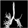
Before and just after ballooning, a chest X-ray was performed in all patients. Baseline and post-ballooning spirometry was performed before and two to three months after ballooning. In addition, the bronchial luminal diameter was estimated and recorded by bronchoscopy. For selected patients, a three dimensional CT scan was obtained (Figure 1).
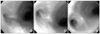
Ballooning was repeated at two to three month intervals and when dyspnea developed (Figure 2). However, if severe bronchomalacia occurred after ballooning, a stent was inserted at the time of the intervention. In addition, if ballooning was ineffective as a result of tight fibrosis, surgical resection and anastomosis were considered.
When stabilized, patients were followed at two to three month intervals with spirometry and chest X-ray. The last FEV1 and FVC were recorded as the post-ballooning spirometric data.
The clinical outcome of patients was considered "successful" if two of following three parameters were met: 1) subjectively improved dyspnea, 2) improved spirometry score, and 3) widened bronchus on follow-up bronchoscopy.
Results
1. Characteristics of patients
Twenty-nine patients were included in this study. The median age of the patients was 28 (range 16~62 year) and most patients were female (n=27, 93.1%). Effective anti-TB medications were started at 4.6±5.9 years (range 0.5~26 years) before the intervention and completed in 23 of the patients. In the remaining six patients, anti-TB drugs were still being taken for at least three months. The duration of anti-TB medication was 10.7±4.5 months (range 6~24 months); isoniazid, rifampin, ethambutol and/ or pyrazinamide were prescribed. No sputum acid fast bacilli (AFB) were detected in patients during the intervention and there were no cases of drug resistant TB.
The involved site was the left main bronchus in 22 patients (75.9%), the right bronchus intermedius in six (20.7%) and the right main in one (3.4%). The baseline forced expiratory volume at one second (FEV1) and the forced vital capacity (FVC) were 66.2±11.9% and 76.1±15.0% of predicted, respectively. The baseline bronchial luminal diameter was 2.9±1.4 mm (range 1~5.5 mm).
The baseline chest X-ray showed complete atelectasis of a pulmonary lobe in 12 patients (41.4%). Complete atelectasis of the left lower lobe was seen in six patients, left upper lobe in three and right upper lobe in three.
2. Clinical outcome
The mean number of ballooning procedures was 2.4 (range 1~8) and the interval between ballooning procedures was 76.2±69.7 days. The Nd-YAG laser was used in six patients who had localized dense fibrosis.
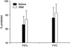
FEV1 and FVC were improved after ballooning (from 66.2±11.9 to 73.5±13.0% predicted, p=0.0004 and from 76.1±15.0 to 83.1±12.6% predicted, p=0.002 respectively, Figure 3). The estimated bronchial diameter was also improved after ballooning compared to the baseline (from 2.9±1.4 mm to 7.2±1.1 mm, p=0.0001).
Pneumothorax developed in two patients (6.9%) who were treated with chest tube insertion. Minor subcutaneous emphysema and pneumomediastinum were observed in three patients (10.3%) who spontaneously recovered. There was no other major complication such as massive hemoptysis, myocardial infarction, or respiratory failure. There was no procedure related death.
3. Subgroup analysis between successful and unsuccessful groups
Among 29 patients, a clinically successful outcome was observed in 16 patients (55.2%). In the remaining 13 patients, whose clinical outcome was unsuccessful, bronchial stenting was performed in nine patients, surgical resection and anastomosis were carried out in one patient and three patients declined further treatment.
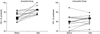
In patients with a successful clinical outcome, FEV1 and FVC were significantly improved after the ballooning compared to baseline (from 71.1±8.1 to 82.8±7.6% predicted, p=0.0005 and from 79.4±13.4 to 90.1±10.9% predicted, p=0.0049 respectively). On the other hand, FEV1 and FVC were not improved after ballooning in the unsuccessful group of patients (from 60.2±13.3 to 63.4±9.2% predicted, p=0.19 and from 72.2±16.4 to 75.6±9.8% predicted, p=0.24 respectively, Figure 4).
In a comparison between the successful and unsuccessful groups, favorable prognostic factors included: higher pre ballooning FEV1 (71.1±8.1 vs. 60.2±13.3, p=0.018), higher post ballooning FEV1 (89.2±7.8 vs. 63.4±9.2, p=0.00004), and absence of complete left upper lobe collapse. However, the clinical outcome did not differ according to age, gender, duration of anti-TB medication, duration between initial TB medication and ballooning, site of bronchial stenosis, bronchial luminal diameter estimated by bronchoscopy, number of ballooning procedures or laser usage. Two patients in whom pneumothorax developed had an unsuccessful clinical outcome. However, the complication rate was not significantly different between the successful and unsuccessful group (18.8% vs. 23.1%).
The clinical outcome was unsuccessful in all eight patients whose pre-ballooning FEV1 was the same as or less than 57% of predicted or had a collapsed left upper lobe of the lung.
Discussion
In this study, bronchial ballooning was successful in about half of the patients with PTBS; the procedure was well tolerated and safe; consistent with previous reports11-15. In addition, we found that the favorable prognostic factors for ballooning in patients with PTBS included: a good baseline FEV1 (more than 57% predicted) and absence of left upper lobe lung collapse.
The mechanism underlying airway stenosis after endobronchial TB may be explained in several ways: destruction of bronchial cartilage by caseation necrosis, cicatric annular stricture due to fibrosis, a mural tuberculoma occluding the bronchial lumen and intramural caseation material5-8. The last two lesions are frequently observed in active endobronchial TB1,2. After effective anti-TB chemotherapy, intramural inflammation and caseation material are replaced by fibrosis, which results in stricture of the bronchial lumen. In addition, the presence of peribronchial inflammation, such as with lymphadenitis, would aggravate fibrosis. Considering the mechanism underlying the development of PTBS, ballooning may be a safe and effective approach to dilatation of the narrowed bronchus by stretching and expanding fibrotic tissue within the bronchial wall.
However, excessive stretching may tear the bronchial wall resulting in pneumothorax, pneumomediastinum and subcutaneous emphysema. During the initial period of time of the trial we applied up to an atmospheric pressure of seven to treat tight fibrosis. However, the result of the excessive high pressures was tearing of the normal bronchial wall rather than stretching of the fibrotic lesion. The current preferred method for localized dense fibrotic lesions is use of the Nd-YAG laser, which is favored for cutting the fibrotic bands, instead of applying excessive high pressure with ballooning.
Previous studies have reported that bronchial stenosis recurred in 30~80% of patients with PTBS who underwent balloon dilatation11,12. Especially when high pressure (up to 16 atmospheric pressures) was applied, the recurrence rate was 80%11. In this study, we applied atmospheric pressures of three and nine out of 29 patients (31.0%) required bronchial stenting for recurrent bronchial stenosis. The recurrence rate in this study was similar to previous reports (27% after 6 months and 36% in after 1 year) that applied atmospheric pressures less than two to five for ballooning12.
After ballooning, remodeling developed at the expanded bronchus. The extent and rate of bronchial remodeling would depend on the fibrotic activity of the expanded bronchus, which depended on intramural or peribronchial inflammation. In this study, bronchial ballooning was performed after a complete course of anti-TB medication when possible. Chronic inflammation due to endobronchial TB could cause deterioration of dilated bronchial patency. In addition, excessive mechanical expansion could aggravate bronchial remodeling by tearing normal bronchial wall tissue. By applying Nd-YAG laser for cutting dense fibrotic lesions and gently expanding normal and less fibrotic tissues, we could decrease the recurrence of bronchial stricture.
In patients with poor baseline FEV1, bronchial ballooning was less effective. As the mechanism of ballooning is mechanical stretching and expanding of the fibrotic bronchial wall, there should be limitations to its application. The ratio of elastic to non-elastic tissue fibers in the bronchial wall is an important factor for successful ballooning. In patients with poor baseline FEV1, the degree of bronchial stenosis would be expected to be more severe, the bronchial wall would be predicted to be more fibrotic, and thus the clinical outcome of ballooning would be expected to be less successful.
There were 12 patients whose baseline X ray showed complete atelectasis of a pulmonary lobe. Complete atelectasis of the left lower lobe was seen in six patients, left upper lobe in three, and right upper lobe in three. All three patients with complete left upper lobe collapse had unsuccessful clinical outcomes. When the left upper lobe collapses, the left pulmonary structures are displaced upward and the left main bronchus is continuously stressed by an upward bending force, which would be expected to aggravate bronchial remodeling.
Although airway stenting is not the focus of this study, the use of metallic mesh stents for patients with PTBS should be chosen with caution. Successful outcomes have been observed in one study16, but the same investigators as well as other groups have reported unfavorable long-term effects of the metallic mesh stent12,17. As PTBS is benign in nature and affects primarily young patients, long term outcome and complications should be carefully considered before intervention.
In summary, bronchial ballooning was successful in about half of the patients with PTBS; it was safe and effective with a more favorable outcome in patients with only marginally reduced baseline pulmonary function and absence of left upper lobe lung collapse. In patients with more advanced bronchial stenosis, more effective modalities, such as laser ablation, bronchial stenting or surgical intervention may be necessary.
 XML Download