Abstract
Lung cancer is one of the most prevalent cancers and it has the highest mortality of all forms of cancers. Although surgery, chemotherapy and radiotherapy are routinely used for the treatment of lung cancer treatment, little progress has been made in the treatment of this condition over the past 20 years. The histological subtype of squamous cell carcinoma (SCC) accounts for approximately 30% of all lung cancer patients. Spontaneous regression of non-small-cell lung cancer (NSCL) is an extremely rare phenomenon. Spontaneous regression of cancer (SR) is defined as a complete or partial, temporary or permanent disappearance of all or at least some the relevant parameters of soundly diagnosed malignant disease without any medical treatment or with treatment that is considered inadequate to produce the resulting regression.
Figures and Tables
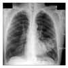
Figure 1
(A) Focal nodular radiopacity and infiltration on left lower lung field. About 8 cm sized lobulating mass in anteromedial basal segment of left lower lung. (B) Subcarinal, both hilar, parabronchial lymphadenopathy.

Figure 2
(A) Squamous cell carcinoma (SCC), moderately differentiated (H&E stain, ×40). (B) Individual cells show hyperchromatic nuclei with keratinization in cytoplasm. Frequent abnormal mitoses are seen (H&E stain, ×100). (C) Abnormal cell clusters in SCC. A keratinizing spindle cell with hyperchromatic nucleus is seen in upper left corner (H&E stain, ×100).

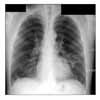
Figure 3
(A) Improved state of left lower lung infiltration. Previously noted lobulated mass in left lower lung show more decreased. (B) Small lymph nodes in parabronchial and A-P window are still remained.

References
1. Yasufuku K, Nakajima T, Motoori K, Sekine Y, Shibuya K, Hiroshima K, et al. Comparison of endobronchial ultrasound, positron emission tomography and CT for lymph node staging of lung cancer. Chest. 2006. 130:710–718.
2. Korean Academy of Tuberculosis and Respiratory Diseases. Respiratory Diseases. 2004. Seoul: Koonja;599–607.
3. Liang HL, Xue CC, Li CG. Regression of squamous cell carcinoma of the lung by Chinese herbal medicine: a case with an 8-year follow-up. Lung Cancer. 2004. 43:355–360.
4. Kappauf H, Gallmeier WM, Wünsch PH, Mittelmeier HO, Birkmann J, Büschel G, et al. Complete spontaneous remission in a patient with metastatic non-small-cell lung cancer. Case report, review of the literature, and discussion of possible biological pathways involved. Ann Oncol. 1997. 8:1031–1039.
5. Cafferata MA, Chiaramondia M, Monetti F, Ardizzoni A. Complete spontaneous remission of non-small-cell lung cancer: a case report. Lung Cancer. 2004. 45:263–266.
6. Hirano S, Nakajima Y, Morino E, Fujikura Y, Mochizuki M, Takeda Y, et al. A case of spontaneous regression of small cell lung cancer with progression of paraneo plastic sensory neuropathy. Lung Cancer. 2007. 58:291–295.
7. Nam SW, Han JY, Kim JI, Park SH, Cho SH, Han NI, et al. Spontaneous regression of a large hepatocellular carcinoma with skull metastasis. J Gastroenterol Hepatol. 2005. 20:488–492.
8. Ikeda M, Okada S, Ueno H, Okusaka T, Kuriyama H. Spontaneous regression of hepatocellular carcinoma with multiple lung metastases: a case report. Jpn J Clin Oncol. 2001. 31:454–458.


 PDF
PDF ePub
ePub Citation
 Print
Print


 XML Download