

 PDF
PDF ePub
ePub Citation
 Print
Print


Abstract
Background
Tsutsugamushi, leptospirosis and hemorrhagic fever with renal syndrome (HFRS) are the prevalent diseases among the acute febrile illnesses in Korea. Pulmonary involvement in the patients with these diseases remains poorly recognized in endemic regions, and this is despite reports of recent outbreaks and epidemic episodes. Pulmonary involvement and a higher CRP level as clinical manifestations show a more severe form of infection. The aim of this study is to analyze the correlation of pulmonary involvement and the CRP level in patients with acute febrile illnesses.
Methods
We retrospectively reviewed the clinical records of 105 patients who were diagnosed with tsutsugamushi, HFRS and leptospirosis from January 2002 to May 2008 in Chuncheon Sacred Heart Hospital. The radiographic images were retrospectively analyzed by two radiologists. We analyzed the pulmonary complications of the patients with these febrile diseases and we checked the CRP level at admission.
Results
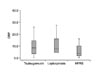
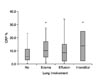
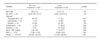
The study included 105 patients who were diagnosed with febrile diseases. Of these patients, 32 patients had hantaan, 10 patients had leptospirosis and 63 patients had tsutsugamushi disease. 42 (40%) patients had pulmonary complications, 20 patients had pulmonary edema, 20 patients had pleural effusion and 2 patients had interstitial pneumonitis. The patients with pulmonary involvement showed a more severe form of infection and a higher CRP level than that of those patients without pulmonary involvement (p=0.0073).
Figures and Tables
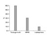
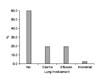
Figure 4
Diagnosis and CRP level. Data are presented as box-plots, where the horizontal line represents the mean.
References
1. Song JW, Lee JE, Kim SH, Kee SH, Park KS, Baek LJ, et al. Seroepidemiologic analysis of acute febrile illness in Korea during 1997-1998. J Bacteriol Virol. 2002. 32:263–267.
2. Munro-Faure AD, Andrew R, Missen GA, Mackay-Dick J. Scrub typhus in Korea. J R Army Med Corps. 1951. 97:227–229.
3. Weil A. Ueber Eile Eligenthumliche, Mit Miltz tumor. Icterus and Nephritis Einhergehende. Acute infectionskrankheit Dtsch Arch Klin Med. 1886. 39:209–232.
4. Martínez García MA, de Diego Damiá A, Menéndez Villanueva R, López Hontagas JL. Pulmonary involvement in leptospirosis. Eur J Clin Microbiol Infect Dis. 2000. 19:471–474.
5. Thammakumpee K, Silpapojakul K, Borrirak B. Lep tospirosis and its pulmonary complications. Respirology. 2005. 10:656–659.
6. Lee WS, Wang FD, Wang LS, Wong WW, Young D, Fung CP, et al. Scrub typhus complicating acute respiratory distress syndrome: a report of two cases. Zhong hua Yi Xue Za Zhi (Taipei). 1995. 56:205–210.
7. Park HK, Jung SJ, Lee SP, Jin SH, Lee KH, Park DC, et al. Clinical study of 27 patients with Tsutsugamushi disease in the Ulsan, Ulchu Area. Korean J Med. 1988. 35:383–389.
8. Jeon KY, Choi YS. Clinical observation of 98 cases of Tsutsugamushi disease (1986-1991). Korean J Med. 1993. 45:177–186.
9. Burke BJ, Searle JF, Mattingly D. Leptospirosis presenting with profuse haemoptysis. Br Med J. 1976. 2:982.
10. Havens WP, Buchar CJ, Raimann HA. Leptospirosis. JAMA. 1941. 116:289.
11. Chiu YC, Liu HH. Report on observation roentgenologic pulmonary changes in 48 cases of leptopirosis. Chin J Radiol. 1959. 7:374–375.
12. Kim WY, Han SY, Kim SK, Suh JE, Joo SA, Kim KM. The 16 cases of leptospirosis in Chuncheon area. Korean J Med. 1986. 30:745–750.
13. Kim HJ, Jung JH, Lee JU, Lee OY, Yang SC, Han DS, et al. Chest X-ray findings and acid-base disturbances in the early phase of Leptospirosis. Korean J Med. 1997. 52:24–31.
14. Padula PJ, Edelstein A, Miguel SD, López NM, Rossi CM, Rabinovich RD. Hantavirus pulmonary syndrome outbreak in Argentina: molecular evidence for person-to-person transmission of Andes virus. Virology. 1998. 241:323–330.
15. Graziano KL, Tempest B. Hantavirus pulmonary syndrome: a zebra worth knowing. Am Fam Physician. 2002. 66:1015–1020.
16. Khan AS, Ksiazek TG, Peters CJ. Hantavirus pulmonary syndrome. Lancet. 1996. 347:739–741.
 XML Download