

 PDF
PDF ePub
ePub Citation
 Print
Print


Abstract
Sarcoidosis is a systemic granulomatous disease that primarily affects the lung and lymphatic system of the body. Since Brincker first noted a statistically significant increase of malignant tumors among sarcoidosis patients, there have been several reports on simultaneously developed sarcoidosis and malignancy. A 30-year-old man was admitted to our hospital because of multiple enlarged mediastinal lymph nodes. The patient had been well until approximately 10 days before admission, when he developed a cough. Chest X-ray and computed tomography (CT) of the chest that were performed at the outpatient department revealed multiple enlarged mediastinal lymph nodes. Cervical lymph node biopsy revealed both non-caseating granuloma and metastatic papillary carcinoma, whereas the mediastinal lymph node showed only non-caseating granuloma. The thyroid gland surgical specimen showed papillary carcinoma. We report here on a case of a 30-year-old man who had sarcoidosis and thyroid cancer, and we include a review of the literature.
Figures and Tables
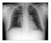
Figure 2
Chest CT shows bilateral hilar and mediastinal lymphadenopathy and wide spread small nodules with a bronchovascular distribution in both lungs.
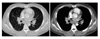
Figure 3
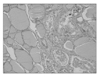
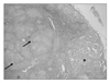
(A) Multiple well-formed small non-caseating granulomas are observed in cervical lymph nodes (short arrows). A multinucleated giant cell is also seen (arrow head) (H&E stain, ×100). (B) Metastatic papillary carcinoma from thyroid is seen in lymph nodes (long arrows) (H&E stain, ×100).
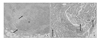
Figure 4
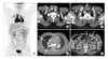
18F-FDG PET/CT reveals multiple lymphadenopathy with FDG hypermetabolism in bilateral hilar & mediastinal (A, D), right supraclavicular (B, arrow) and mesenteric areas (E). A thyroid nodule located in left lower thyroid gland shows no demonstrable FDG hypermetabolism (B, arrow head). Perithyroidal lymph node with mild FDG hyperactivity (C, arrow) has proved a metastatic and sarcoidosis node. Small peribronchovascular pulmonary nodules are seen in right upper and right lower lobes with faint increased FDG uptake (D).
References
1. Statement on sarcoidosis. Joint Statement of the American Thoracic Society (ATS), the European Respiratory Society (ERS) and the World Association of Sarcoidosis and Other Granulomatous Disorders (WASOG) adopt ed by the ATS Board of Directors and by the ERS Executive Committee, February 1999. Am J Respir Crit Care Med. 1999. 160:736–755.
2. Iannuzzi MC, Rybicki BA, Teirstein AS. Sarcoidosis. N Engl J Med. 2007. 357:2153–2165.
3. Nunes H, Soler P, Valeyre D. Pulmonary sarcoidosis. Allergy. 2005. 60:565–582.
4. Baughman RP, Lower EE, du Bois RM. Sarcoidosis. Lancet. 2003. 361:1111–1118.
5. Brincker H. Sarcoid reactions and sarcoidosis in Hodgkin's disease and other malignant lymphomata. Br J Cancer. 1972. 26:120–123.
6. Brincker H, Wilbek E. The incidence of malignant tumors in patients with respiratory sarcoidosis. Br J Cancer. 1974. 29:247–251.
7. Brincker H. Sarcoidosis and malignancy. Chest. 1995. 108:1472–1474.
8. Reich JM, Mullooly JP, Johnson RE. Linkage analysis of malignancy-associated sarcoidosis. Chest. 1995. 107:605–613.
9. Askling J, Grunewald J, Eklund A, Hillerdal G, Ekbom A. Increased risk for cancer following sarcoidosis. Am J Respir Crit Care Med. 1999. 160:1668–1672.
10. Cohen PR, Kurzrock R. Sarcoidosis and malignancy. Clin Dermatol. 2007. 25:326–333.
11. Pandha HS, Griffiths H, Waxman J. Sarcoidosis and cancer. Clin Oncol (R Coll Radiol). 1995. 7:277–278.
 XML Download