

 PDF
PDF ePub
ePub Citation
 Print
Print


Abstract
Exogenous lipoid pneumonia (ELP) is a chronic inflammatory reaction of the lungs resulting from the aspiration of vegetable, animal or mineral oils. Squalene, is a derivative of shark liver oil that is taken as a traditional remedy in some Asian countries, and is used widely also in cosmetics. Similar to the symptoms in most cases of oil aspiration, the symptoms of squalene-induced lipoid pneumonia are either absent or nonspecific. Hence, the disease is generally detected incidentally. Although many cases with predisposing factors have been reported, ELP with achalasia is quite rare. We report a 47-year old woman with achalasia who developed ELP after ingesting squalene. The patient was treated successfully by supportive care and surgical treatment of the achalasia.
Figures and Tables
Figure 1
(A) On admission, chest radiography shows ground-glass appearance on right upper and lower lung field. (B) 6 month later, chest radiography shows improvement.
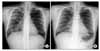
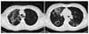
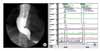
Figure 2
High resolution computed tomography scan of the chest shows multifocal GGA and nodule with crazy paving appearance on right upper lobe and esophageal dilatation.
References
1. Spickard A 3rd, Hirschmann JV. Exogenous lipoid pneumonia. Arch Intern Med. 1994. 154:686–692.
2. Laughlen GF. Studies on pneumonia following nasopharyngeal injection of oil. Am J Pathol. 1925. 1:407–414.
3. Gondouin A, Manzoni P, Ranfaing E, Brun J, Cadranel J, Sadoun D, et al. Exogenous lipid pneumonia: a retrospective multicentre study of 44 cases in France. Eur Respir J. 1996. 9:1463–1469.
4. Choi HS, Kwag HJ, Chae SW, Lim SY, Lim SY. Severe exogenous lipoid pneumonia following ingestion of large dose squalene: successful treatment with steroid. Tuberc Respir Dis. 2006. 60:235–238.
5. Hyun JG, Rhee JH. Clinical investigation of lipoid pneumonia in adult. Tuberc Respir Dis. 1996. 43:965–975.
6. Lee JS, Ju HD. A case of lipoid pneumonia induced by aspiration of shark liver oil. Tuberc Respir Dis. 1994. 41:670–675.
7. Park HP, Kwon KY, Choi WI. Lipoid pneumonia in Korea: a case report and review of the literature of Korean cases. Respir Med Extra. 2007. 3:39–43.
8. Jeong KJ, Kim YE, Lim GJ, Suh KD, Kim JD, Lee JH, et al. A case of lipoid pneumonia after ingestion of green perilla oil. Tuberc Respir Dis. 1999. 47:123–126.
9. Wright BA, Jeffrey PH. Lipoid pneumonia. Semin Respir Infect. 1990. 5:314–321.
10. Hughes RL, Freilich RA, Bytell DE, Craig RM, Moran JM. Clinical conference in pulmonary disease: aspiration and occult esophageal disorders. Chest. 1981. 80:489–495.
11. Laurent F, Philippe JC, Vergier B, Granger-Veron B, Darpeix B, Vergeret J, et al. Exogenous lipoid pneumonia: HRCT, MR, and pathologic findings. Eur Radiol. 1999. 9:1190–1196.
12. Rossi SE, Erasmus JJ, Volpacchio M, Franquet T, Castiglioni T, McAdams HP. "Crazy-paving" pattern at thin-section CT of the lung: radiologic-pathologic overview. Radiographics. 2003. 23:1509–1519.
 XML Download