Abstract
Systemic lupus erythematosus (SLE) is a multisystem disorder where the etiology is not clearly known. Symptomatic chronic interstitial pneumonitis is an uncommon manifestation, with a reported prevalence of 3~13%. Achalasia is rare disease that presents with failure in the relaxation of the esophagus sphincter. A 22-year-old woman was admitted to our hospital because of fever, cough and dyspnea. The patient had a history of pericardial effusion and Raynaud's phenomenon. The results of laboratory tests indicated the presence of lymphopenia and included positive antibody tests for antinuclear antibody and anti Sm antibody. A chest X-ray demonstrated the presence of peribronchial infiltration on both lung fields. A Chest CT image showed interlobar septal thickening, ground-glass opacity and a honeycomb appearance in both lung fields and esophageal dilatation with air fluid level. An esophagogram showed the presence of dilated esophagus ends that represented the non-relaxed lower esophageal sphincter. Manometry demonstrated incomplete sphincter relaxation. The case was diagnosed as systemic lupus erythematosus associated with interstitial pneumonia and achalasia.
Figures and Tables
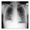
Figure 1
Chest PA shows diffuse reticularities with peribronchial infiltration on both lower lung fields.

References
1. Lopez-Liuchi JV, Kraytem A, Uldry PY. Oesophageal achalasia secondary to pleural mesothelioma. J R Soc Med. 1999. 92:24–25.
2. Kim JS, Lee KS, Koh EM, Kim SY, Chung MP, Han J. Thoracic involvement of systemic lupus erythematosus: clinical, pathologic, and radiologic findings. J Comput Assist Tomogr. 2000. 24:9–18.
3. Wiedemann HP, Matthay RA. Pulmonary manifestations of the collagen vascular diseases. Clin Chest Med. 1989. 10:677–722.
4. Feldman M. Esophageal achalasia syndromes. Am J Med Sci. 1988. 295:60–81.
5. Goldschmiedt M, Peterson WL, Spielberger R, Lee EL, Kurtz SF, Feldman M. Esophageal achalasia secondary to mesothelioma. Dig Dis Sci. 1989. 34:1285–1289.
6. Park SK, Kwon HC, Koh IY, Kim KH, Lee CM, Lee SW, et al. A case of systemic sclerosis associated with gastric cancer. Korean J Med. 2005. 69:326–329.
7. Vaezi MF, Richter JE. Diagnosis and management of achalasia. American College of Gastroenterology Practice Parameter Committee. Am J Gastroenterol. 1999. 94:3406–3412.
8. Chung YH, Rhee PL, Jung HC, Song YW, Lee HS, Yoon YB, et al. Correlation of esophageal symptoms with esophageal motility in patients with progressive systemic sclerosis. Korean J Gastroenterol. 1988. 20:519–524.
9. Zamost BJ, Hirschberg J, Ippoliti AF, Furst DE, Clements PJ, Weinstein WM. Esophagitis in scleroderma: prevalence and risk factors. Gastroenterology. 1987. 92:421–428.


 PDF
PDF ePub
ePub Citation
 Print
Print


 XML Download