

 PDF
PDF ePub
ePub Citation
 Print
Print


Abstract
Neurilemmomas are benign nerve sheath tumors derived from Schwann cells that rarely occur in the chest wall. Neurilemmomas of the chest wall are usually solitary lesions that bulge toward the pleural cavity. Neurilemmomas are confirmed histologically based on the presence of Verocay bodies, Antoni A and Antoni B tissue patterns and S-100 protein. Bilateral neurilemmomas in the chest wall are extremely rare, as are those that grow in the subcutaneous tissue but not the pleural area. We report here a case of bilateral chest wall neurilemmomas in which the tumors bulged out to the skin and were palpable.
Figures and Tables
Figure 1
Chest PA and left lateral radiographic findings of 62 year old patients who had already known pulmonary nodules. Faint mass like density are found at left 4th intercostal space (white arrow) and soft tissue swelling in right lower chest (black arrow).
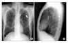
Figure 2
Chest computed tomography of the presented patients show soft and oval shape homogenous low density mass at the intercostal area, protruding to lung (A). Lower right chest area same density mass are detected in the pleura which is protruded toward subcutaneous tissue (B).
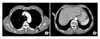
Figure 3
Specimens from the patient by surgical extirpation are well encapsulated about 2 cm sized single mass from left chest wall (A), and conglobulated five ovoid soft masses from right, 3 cm, the largest one (B).
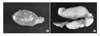
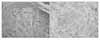
Figure 4
Microscopic examination from the sectioned specimen, tumor was composed two parts, the compact area (Antoni type A) which was condensed spindle cell clusters and less compact area (Antoni type B) which was less condense and degenerated area. The hyalinized acellular area and the rows of parallel nuclei were specific findings of Verocay body (A) (H&H stain, ×100). Immunohistochemical staining showed positive (brown color) for S-100 protein which is stained melanoma and neuron cells (B) (×100).
References
1. Weiss SW, Goldblum JR. Weiss SW, Goldblum JR, Enzinger FM, editors. Schwannoma (neurilemmoma). Enzinger and Weiss's soft tissue tumor. 2001. 4th ed. St. Louis: Mosby;1146–1672.
2. Kim CR, Moon DC, Kwon KS, Chung TA. Neurilemmoma occurring in the skin and lung. Ann Dermatol. 1990. 2:121–125.
3. McClenathan JH, Bloom RJ. Peripheral tumors of the intercostal nerves. Ann Thorac Surg. 2004. 78:713–714.
4. Sakurai H, Hada M, Mitsui T, Ashizawa I. Extrathoracic neurilemmoma of the lateral chest wall mimicking a subcutaneous tumor: report of a case. Ann Thorac Cardiovasc Surg. 2006. 12:133–136.
5. Kim DY, Cho CH, Ahn CM, Sohn HY, Lee DY, Yoon JH. A Case of benign solitary schwannoma of the chest wall. Korean J Intern Med. 1987. 33:119–124.
6. Lee TY, Park JS, Sung YR, Kim WS, Lee JK, Park MK, et al. A case report of neurilemmoma of the chest wall. Tuberc Respir Dis. 1997. 44:649–654.
7. Koh YH, Kim MI, Han MS, Yoo JH, Kang HM. A case of neurilemmoma of the chest wall. Tuberc Respir Dis. 1999. 46:580–585.
8. Ahn CM, Lee HB, Lee YC, Rhee YK. Two cases of intrabronchial neurilemmoma. Tuberc Respir Dis. 2000. 49:225–230.
9. Sakai F, Sone S, Kiyono K, Maruyama A, Ueda H, Aoki J, et al. Intrathoracic neurogenic tumors: MR-pathologic correlation. AJR Am J Roentgenol. 1992. 159:279–283.
10. Gay RE, Gay S, Jones RE Jr. Histological and immunohistological identification of collagens in basement membranes of Schwann cells of neurofibromas. Am J Dermatopathol. 1983. 5:317–325.
11. Evans KG, Miller RR, Muller NL, Nelems B. Chest-wall tumours. Can J Surg. 1990. 33:229–232.
 XML Download