

 PDF
PDF ePub
ePub Citation
 Print
Print


Introduction
Cigarette smoking is the major risk factor in 80~90% of patients with chronic obstructive pulmonary disease (COPD)1. However, only 10~20% of cigarette smokers develop clinically significant airway obstruction, suggesting that genetic factors may play an important role in determining susceptibility to COPD2-4. This genetic susceptibility may result from functional polymorphisms of genes involved in antiproteolysis, xenobiotic metabolism, the inflammatory response to cigarette smoke, and mucociliary clearance3,4.
Human α1-antitrypsin (AAT) is a 54 kDa glycoprotein encoded by SERPINA1, which is part of a serine proteinase inhibitor gene cluster that includes corticosteroid binding globulin (SERPINA6), protein C inhibitor (SERPINA5), and alpha 1-antichymotrypsin (SERPINA3)5. AAT is produced by hepatocytes and mononuclear phagocytes, and functions as a major inhibitor of neutrophil elastase, an ominous protease capable of destroying most components of the lung matrix6. An inherited severe deficiency of AAT increases the risk of COPD, particularly in smokers7,8.
The SERPINA1 gene is located on chromosome 14q32, and more than 100 genetic variants have been identified. The initially discovered AAT variants were named based on electrophoretic migration velocity, as follows: F (fast), M (medium), S (slow), or Z (very slow)9,10. The most common variants of AAT are the M variants, which consist of at least the following four subtypes: M1Val, M1Ala, M2 and M3. The four subtypes differ in their amino acid composition at codons 101, 273 and 376 as follows: M1Val, Arg101-Val213-Glu376; M1Ala, Arg101-Ala213-Glu376; M2, His101-Val213-Asp376; and M3, Arg101-Val213-Asp376 7-11.
The M variants are characterized by normal plasma AAT levels and are thought to be unrelated to any disease 7,12. However, Gaillard et al.13,14 reported that the M1Ala and M2 alleles had a significantly lower elastase inhibitory capacity compared to the M1Val allele. In addition, a number of studies have observed an increased prevalence of the M1Ala and M2 allele in asthmatics13-15. These results suggest that the M1Ala and M2 alleles may play a role in the inflammatory reaction and/or the elastase-antielastase balance, which are important in the pathogenesis of COPD. We conducted a case-control study to evaluate the potential association between SERPINA1 genotypes (M1Val, M1Ala, S and Z) and the risk of COPD.
Materials and Methods
1. Study population
The patient group consisted of 89 male patients who were diagnosed with COPD at the Kyungpook National University Hospital according to the criteria established by the NHLBI/WHO Global Initiative for COPD (GOLD)16. The inclusion criteria for COPD were as follows: chronic respiratory symptoms and signs such as cough and dyspnea; post-bronchodilator FEV1<80% of the predicted value; FEV1/FVC<70%; and FEV1 reversibility after inhalation of 200 µg salbutamol<12% of the pre-bronchodilator FEV1. The severity of COPD was classified by the guidelines of the GOLD in terms of the percentage predicted FEV1, as follows: mild (>80%), moderate (50∼80%), severe (30∼50%), or very severe (<30%). Control subjects (n=112) were selected from a pool of healthy men who visited the general health check-up center. The enrollment criteria for the controls were as follows: male gender, age>45 years, no known disease and no history of any disease, and no airflow limitation. All of the cases and the controls were ethnic Koreans that resided in Daegu City or in the surrounding regions. A trained interviewer completed detailed questionnaires for each patient and each control subject. This study was approved by the Institutional Review Board of the Kyungpook National University Hospital, and written informed consent was obtained from each participant.
2. Genotyping
Genomic DNA was extracted from peripheral blood lymphocytes by proteinase K digestion and phenol/chloroform extraction. Genotypes were determined by PCR-RFLP assay as described previously15,17. The PCR primers for the Ala213Val (M1), Arg101His (M2), Glu264Val (S) and Glu342Lys (Z) variants were 5'-CCCACCTTCCCCTC TCTCCAGGCAAATGGG-3' (forward) and 5'-GGGCCTCA GTCCCAACATGGCTAAGAGGTG-3' (reverse); 5'-GCAGG ACAATGCCGTCTTCTGTCTC-3' (forward) and 5'-CCACTA GCTTCAGGCCCTCGCTGAG-3' (reverse); 5'-TGAGGGG AAACTACAGCACCTCG-3' (forward) and 5'-AGGTGTGG CAGCTTCTTGGTCA-3' (reverse); and 5'-ATAAGGCTGT GCTGACCATCGTC-3' (forward) and 5'-TTGGGTGGGAT TCACCACTTTTC-3' (reverse), respectively. The PCR reactions were performed in a total volume of 20 µl that contained 200 ng genomic DNA, 10 pM of each primer, 4 mM dNTPs, 10 mM Tris-HCl (pH 8.3), 50mM KCl, 1.5 mM MgCl2, and 1 unit of Taq polymerase (Takara Shuzo Co., Otsu, Shiga, Japan). The PCR cycle conditions consisted of an initial denaturation step at 94℃ for 5 min followed by 35 cycles for 30 s at 94℃, 20 s at 67℃ for the M1 and M2 variants, and 55℃ for the S and Z variants, 30 s at 72℃, and a final elongation step at 72℃ for 10 min.
The restriction enzyme, BstEII (New England BioLabs, Beverly, MA, USA), was used to distinguish the M1 variant in which the gain of a BstEII restriction site in addition to a restriction site at codon 288 occurs in the Val allele. Thus, the Val allele yields three bands (228, 83 and 49 bp) and the Ala allele yields two bands (311 and 49 bp). The restriction enzyme RsaI (New England BioLabs, Beverly, MA, USA) was used to distinguish the M2 variants in which the loss of the RsaI restriction site occurs in the His allele. The Arg allele yields two bands (383 and 79 bp) and the His allele has only one band representing the entire 462 bp fragment. The restriction enzyme, TaqI (New England BioLabs, Beverly, MA, USA), was used to distinguish the S and Z variations. Using the primers for the S variant, the S allele has a band at 121 bp whereas the M allele has a band at 157 bp. With the primers for the Z variant, the Z allele has a band at 179 bp and the M allele has a band at 157 bp. Five microliters of the PCR products were digested overnight with 5 U BstEII at 60℃, 5 U RsaI at 37℃ or 10 U TaqI at 65℃. The digestion products were separated on 2.5% agarose gel for the M1 and M2 variants and on 8% acrylamide gel for the S and Z variants.
3. Statistical analysis
The cases and controls were compared using the Student's t-test for continuous variables and a χ2 test for categorical variables. Unconditional logistic regression analyses were used to calculate the odds ratios (ORs) and 95% confidence intervals (CIs), with adjustment for possible confounders (age and pack-years of smoking as continuous variables). In addition to the overall association analysis, we performed a stratified analysis according to age (median age,<62 years/≥ 62 years), smoking status and severity of COPD (GOLD I-II/GOLD III-IV) to further explore the association between genotypes and the risk of COPD in each stratum. The homogeneity test was done to compare the difference between genotype/diplotype-related ORs of different groups. p-values from the analyses results<0.05 were considered as statistically significant. All of the analyses were performed using Statistical Analysis Software for Windows, version 9.1.3 (SAS Institute, Cary, NC, USA).
Results
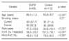
The baseline characteristics of the cases and controls enrolled in this study are shown in Table 1. There was a significant difference in the mean age between the cases and controls (65.4±7.2 vs. 60.8±8.7 years, p<0.001). In addition, the number of pack-years in the smokers was significantly higher in the cases than in the controls (47.2±23.8 vs. 35.8±15.4 pack-years; p<0.001). These differences were controlled for in the subsequent multivariate analyses. The FEV1 and FEV1/FVC ratio were significantly lower in the case group than in the controls (both, p<0.001).
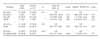
The distributions of SERPINA1 genotypes among the cases and controls are shown in Table 2. The S and Z alleles were not observed in the cases and controls. The frequency of the M1Val, M1Ala, and M2 alleles among the healthy controls were 0.83, 0.005, and 0.17, respectively, which were similar to the healthy Koreans (0.81, 0.003, and 0.19, respectively) in our previous study18, but significantly different from Caucasians (0.44∼0.49, 0.20∼0.23, and 0.14∼0.19, respectively)10. The M1Val allele was significantly less frequent in the cases than in the controls (73.6% vs. 82.7%, p=0.03). The M1Val/M1Val genotype was less frequent in the cases (53.9%) than in the controls (69.2%), whereas the M1Val/M1Ala, M1Val/M2, and M2/M2 genotypes were more frequent in the cases (3.4, 36.0, and 6.7%, respectively) than in the controls (1.0, 26.0, and 3.8%, respectively). These findings suggest that the M1Val/M1Ala, M1Val/M2 and M2/M2 genotypes, which carry the M1Ala or M2 alleles, might be risk genotypes for COPD. Compared with the M1Val/M1Val genotype, the M1Val/M1Ala, M1Val/M2 or M2/M2 genotypes were associated with a significantly increased risk of COPD (adjusted OR: 1.86, 95% CI: 1.02∼3.41, p=0.04). To examine the effect of the M2 allele on the risk of COPD, we combined the M1Val/M2 genotype with the M2/M2 genotype into one susceptible group and compared it with the M1Val/M1Ala genotype. Individuals with at least one M2 allele were at a borderline significantly increased risk of COPD compared with those with the M1Val/M1Val genotype (adjusted OR: 1.77, 95% CI: 0.96∼3.27, p=0.07).
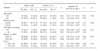
The association between the M2 allele and the risk of COPD was further examined after stratifying the subjects according to age, smoking status, COPD severity and diffusing capacity of the lung for carbon monoxide (DLCO). When stratified by median age, the effect of the combined M1Val/M2 and M2/M2 genotype on the risk of COPD was more pronounced in the subgroup <64 years than in the subgroup ≥64 years (adjusted OR: 3.09, 95% CI: 1.16∼8.21, p=0.02 versus adjusted OR: 1.24, 95% CI: 0.56∼2.76, p=0.60; p in test for homogeneity [pH]=0.04; Table 3). When the subjects were stratified according to the smoking status, the effect of the combined M1Val/M2 and M2/M2 genotype on the risk of COPD was similar in current and former smokers (adjusted OR: 1.80, 95% CI: 0.86∼3.75; and adjusted OR: 1.62, 95% CI: 0.57∼4.63, respectively, pH=0.80). However, when the subjects were dichotomized by median pack-years of smoking, the combined M1Val/M2 and M2/M2 genotype was associated with a borderline significantly increased risk of COPD in lighter smokers (adjusted OR: 2.69, 95% CI: 0.96∼7.52, p=0.06), whereas there was no significant association in heavier smokers (adjusted OR: 1.60, 95% CI: 0.72∼3.53, p=0.25). When the COPD cases were categorized based on disease severity, the effect of the combined M1Val/M2 and M2/M2 genotype on the risk of COPD was similar in individuals with mild-to-moderate COPD (adjusted OR: 1.50, 95% CI: 0.72∼3.10) and those with severe COPD (adjusted OR: 1.91, 95% CI: 0.86∼4.25, pH=0.54). When the COPD cases were categorized based on the DLCO, the combined M1Val/M2 and M2/M2 genotype was associated with a borderline significantly increased risk of COPD in individuals with a DLCO less than 70% of the predicted value (adjusted OR: 2.25, 95% CI: 0.95 ∼5.35, p=0.07), whereas there was no significant association in those with a DLCO ≥70% of the predicted value (adjusted OR: 1.47, 95% CI: 0.73∼2.99, p=0.28).
Discussion
We investigated the association between the SERPINA1 genotypes and the risk of COPD in a Korean population. The M1Val allele was significantly less frequent in the COPD cases than in the controls. Individuals with the M2 or M1Ala allele were at a significantly increased risk of COPD compared to those with the M1Val/M1Val genotype. The association of the SERPINA1 genotypes with the risk of COPD was more evident in the subgroup <64 years. These results suggest that the SERPINA1 genotypes contribute to genetic susceptibility to COPD in Koreans.
In the present study, the M2 allele had a more pronounced effect on the risk of COPD in the subgroup <64 years than in the subgroup ≥64 years, which is consistent with the notation that genetic factors can play a major role in the early onset of disease19,20. Individuals with aberrant AAT activity may be prone to developing COPD at a younger age, thus the association would be more clearly observed in younger patients with COPD. Another interesting finding of this study was that the effect of the M2 allele on the risk of COPD was significant in lighter smokers, yet not significant in heavier smokers. This finding is also biologically plausible, as genetic effect on the risk may be smaller at high levels of tobacco exposure when environmental influences may overpower any genetic predisposition21-23.
The mechanism responsible for the association of the M2 allele with the risk of COPD remains to be elucidated. However, it has been observed that the M2 variant has lower elastase inhibitory capacity compared to the M1Val variant13,14. Therefore, it is possible that individuals with the M2 variant have lower elastase inhibitory capacity, and are thus prone to develop COPD. Another possible mechanism is that the M2 allele may be in linkage disequilibrium with other functional polymorphisms in the SERPINA1 gene (not the Z or S mutations) or in neighboring genes. Because the SERPINA1 gene is located within a cluster of serine protease inhibitor genes that include SERPINA6, SERPINA5, SERPINA3, and Kallistatin24,25, the observed association between the M2 allele and COPD risk may be resulted from linkage disequilibrium with polymorphisms of these adjacent genes.
Several studies have investigated the association between AAT variants and the risk of COPD7,8,26. In contrast to the finding in the present study, the M2 variant was not associated with the risk of COPD in these previous studies. This discrepancy may be due to the different ethnicity of the study populations. The allele and genotype frequencies of the SERPINA1 polymorphisms, as well as the LD status with other variants in neighboring genes, vary greatly between ethnic groups. Therefore, the genetic effects of SERPINA1 polymorphisms may be different in different ethnic populations. The discrepancy may arise from differences in the methods of assigning AAT status. In the previous studies7,8,26, the AAT phenotype was only determined by isoelectric focusing of serum and the serum AAT levels without measuring functional activity of the AAT protein. Although the M2 variant does not affect serum AAT levels, it may alter the functional activity of the AAT protein13,14, and thus predispose to the development of COPD.
In conclusion, the M2 allele of the SERPINA1 gene was significantly associated with the risk of COPD in Koreans. The effect of the M2 allele on the risk of COPD was more pronounced in the subgroup<64 years. These results suggest that SERPINA1 polymorphisms may contribute to a genetic predisposition for COPD. However, it is possible that our findings are attributable to chance because of the relatively small numbers of cases. Therefore, additional studies with larger sample sizes will be required to confirm our findings. Moreover, further study is needed to define a functional role of the M1 allele in the development of COPD.
 XML Download