

 PDF
PDF ePub
ePub Citation
 Print
Print


Introduction
Interest in the role of infection in asthma reemerged during the 1990s, when the importance of viral infections as precipitants of the majority of asthma exacerbations was demonstrated1. Assertions that bacterial infections may have a role in the pathogenesis of asthma-both acute and chronic-are much more controversial2. Of bacterial respiratory pathogens, the atypical bacteria M. pneumoniae is most commonly implicated in each of these contexts.
This article will discuss in brief the biological mechanism of asthma, general feature of M. pneumoniae and the laboratory tests used for diagnosis of this bacterium. Also it reviews the evidence for an association between M. pneumoniae and the pathogenesis of asthma. The possible role of antibacterial therapy in the management of asthma is also discussed.
Asthma
Asthma is best described as a chronic disease that involves inflammation of the pulmonary airways and bronchial hyperresponsiveness that results in the clinical expression of a lower airway obstruction that usually is reversible3. Asthma appears to involve multiple genes that interact with each other and the environment. Twin studied had found a heritability estimate ranging from 36~87%4. The basis of clinical and laboratory findings, asthma has traditionally been divided into atopic (extrinsic) and nonatopic (intrinsic) subgroups. The nonatopic typically has its onset in adulthood, has a more severe clinical course than atopic asthma and is not closely associated with family history of the disease5. Different aetiological mechanisms have been proposed to be involved in the development of these two subgroups, and, in particular, the aetiology of nonatopic, adult-onset asthma remains largely enigmatic6.
Airway remodeling in asthma
Airway remodeling is a fundamental feature of asthma. Disruption and altered function of the epithelium in asthmatics have been proposed as a cause of stimuli that lead to restructuring of the airway wall, as a response to injury. Epithelial cells in asthmatics have been found to have a reduced capacity to repair the epithelial layer following injury and an enhanced capacity to produce proinflammatory and profibrogenic cytokines7. Cytokines play a central role in the coordination and sustenance of the inflammation of the airways in asthma. They are not only involved in maintaining the chronic inflammatory process, but also appear to be able to initiate it8. In this system of complex interactions, the ultimate causes, besides genetic defects, which are responsible for the disturbance of the cytokine balance, and their relative importance, are understandably difficult to determine. However, the possible role of a persistent infection here cannot be ignored6.
Mycoplasma pneumoniae: general features
Mycoplasmas represent the smallest free-living microorganisms and are commonly found in plants, animals, and humans. Because they lack a cell wall, they are resistant to beta-lactam antibiotics, stain poorly with Gram stain, and display marked cellular pleomorphism8. Currently, 16 species isolated from humans are known. In addition, animal mycoplasmas have been detected occasionally in humans, particularly in immunodeficient patients. Out of the 16 species, six are known to cause diseases: M. fermentans, M. hominis, M. genitalium, M. pneumoniae, Ureoplasma urealyticum, and U. parvum. M. pneumoniae is the most important and the most well-known9.
When mycoplasmas colonize, most commonly at mucosal surfaces, chronic inflammatory diseases of the respiratory tract, urogenital tract, and joints often occur. In order for colonization and infection to occur, there must be a strong adhesion of the mycoplasma organism to host cells. This complex process of adhesion requires many accessory proteins8. The major adhesin is a 170-kilodalton (kDa) protein, named P1, important for pathogenesis, being one of the main antigens, with specific antibody production by the host. Cytoadhesion protects mycoplasmas from mucociliary clearance12 and affects its integrity in several ways9.
The loss of ciliary motility results from the production of hydrogen peroxide and superoxide, which injure the epithelia, their cilia, and erythrocyte membranes and provokes in vitro hemolysis, alteration of the erythrocyte antigen and cold agglutinins, which agglutinate erythrocytes in vitro at 4℃. These cold agglutinins are found in the serum of over 50% of patients developing M. pneumoniae pneumonia9. In addition, the organism's structure contributes to the inhibition of ciliary movement because its filamentous end allows it to slip between cilia within the respiratory epithelium10. Once adhesion is achieved, the incubation period lasts for 1 to 3 weeks. This long incubation period is what makes M. pneumoniae spread slowly from person to person8.
Detection of Mycoplasma pneumoniae
Investigation of the potential association between M. pneumoniae and asthma is greatly hampered by the lack of standardized, sensitive, and specific methods for the detection of this atypical respiratory pathogen11.
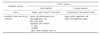
M. pneumoniae infections are usually documented by serology using a complement fixation test (CFT) or by culture through isolation from throat, nasopharynx and pleural fluid. Both these tests have limitations in terms of sensitivity and specificity. Serological tests are often hampered by interspecies cross-reactions, whereas culture is insensitive, time-consuming, and hard to achieve for clinical samples, limiting its usefulness for routine purposes12. Therefore more sensitive and specific tests are needed for exact diagnosis of M. pneumoniae-associated infections. At present, only a few laboratories offer more specific and sensitive techniques using two different approaches. One is direct detection of antigen and the other is to use a method capable of detecting anti- M. pneumoniae antibodies13,14. Direct detection tests such as antigen detection, hybridization with DNA probes, and, more recently, PCR were developed. DNA probes and antigen-detecting enzyme-linked immunosorbent assay (ELISA) have been shown to be effective substitutes for culture. However, cross-reactivity between M. pneumoniae and M. genitallium has been observed with both the methods. Furthermore, detection by DNA probe lacks sensitivity15. Presently, PCR for detection of M. pneumoniae has been recommended for better detection. Different primer sets for the detection of M. pneumoniae have been described; the targets are gene coding for the P1 adhesin protein, the 16SrRNA gene, a DNA sequence specific for M. pneumoniae selected from genomic library, and the gene coding elongation factor Tu16. PCR has opened the possibility to improve the diagnosis of M. pneumoniae infections. It seems to be the best method of detection. Advantages include promptness (hours), early diagnosis (does not depend on serological conversion or agent growth), and detection (small quantities) in conventionally anomalous sites (blood and airway secretions, liquor). Sensitivity is theoretically very high; it is able to identify a single organism when purified DNA is used and it does not require viable organisms9. Contamination is the greatest problem for diagnosis by PCR and is responsible for a great number of wrong diagnoses. Various methods of diagnosis are summarized in Table 1.
Epidemiology for association of Mycoplasma pneumoniae and asthma
M. pneumoniae infections have been associated with asthma. M. pneumoniae infection may be associated with longlasting dry cough, and an association with asthma symptoms has been suggested18. The main problem that hampers the full understanding of the possible association between M. pneumoniae infection and asthma is the lack of standardized, sensitive, and specific diagnostic methods19.
Information linking M. pneumoniae infection to acute and chronic asthma exacerbations has been gathered both for children and adults.
M. pneumoniae in asthmatic children
A relationship between acute infection with M. pneumoniae and acute asthma exacerbations in children has been sought in several controlled and uncontrolled studies. The vast majority of studies were concordant in finding an association between M. pneumoniae infection and asthma exacerbations. Rates of identification varied between 5% and 22.5% of asthma episodes for M. pneumoniae19,20. These findings suggest a relationship between childhood asthma and acute M. pneumoniae infection.
In children, with wheezing, the incidence of acute M. pneumoniae infections increases with age and occurs mainly after 5 years of age. Esposito et al.21 studied 225 children with an acute episode of wheezing, 16 of whom had Mycoplasma infection as determined by serology and/or PCR on nasopharyngeal secretions, and compared them to 8 asymptomatic children with M. pneumoniae and 8 uninfected controls. Children with wheezing and acute M. pneumoniae infection had a statistically significant increase in interleukin-5 (IL-5) compared to children with M. pneumoniae who were asymptomatic and to the controls without wheezing. Those authors therefore proposed that M. pneumoniae might trigger the wheezing process by means of IL-5 secretion in persons who are genetically predisposed or are otherwise susceptible. This seems plausible since IL-5 is the cytokine that has been shown to be essential for development of airway hyperresponsiveness in association with infection caused by respiratory syncytial virus22.
In addition to acute exacerbations, the role of M. pneumoniae in the pathogenesis of chronic asthma has also been extensively investigated. Accumulating evidence from sero-epidemiological studies has shown that many asthmatics have elevated antibody levels and M. pneumoniae23. It has been proposed that both pathogens cause occult chronic lower airway inflammation. Consequently, an association between M. pneumoniae and chronic asthma has been hypothesized2.
Trials addressing the possible relationship between M. pneumoniae and persistent asthma in children have found positive. Gil et al.24 reported that throat cultures for M. pneumoniae were positive in 24.7% of children and adults with asthma exacerbation, compared with 5.7% of healthy controls. Moreover, there is strong evidence suggesting that serologic evidence of M. pneumoniae in children hospitalized with acute exacerbations of asthma is associated with persistent asthma symptoms25.
M. pneumoniae in asthmatic adults
Studies employing serology, culture and molecular biology to identify acute M. pneumoniae infection have been carried out in adult patients presenting with asthma exacerbations26,27. Evidence of M. pneumoniae infection was found in most, but not all of these studies. Lieberman et al.28 found that M. pneumoniae was associated with hospitalization for acute exacerbation of bronchial asthma. Overall, available studies in adults and children suggest that acute M. pneumoniae infection may play a significant role in asthma exacerbations.
A large number of controlled and uncontrolled trials have investigated the role of M. pneumoniae in adult patients with chronic asthma29,30. Once again, serology was the most commonly employed diagnostic method. Most trials did observe an association between M. pneumoniae infection and chronic asthma. Kraft et al.29 have shown that asthmatic patients exhibit evidence of M. pneumoniae airway colonization/infection with significantly greater frequency than non-asthmatic subjects.
Martin et al.23 reported one of the most comprehensive evaluations of the role of M. pneumoniae infections in patients with chronic asthma. In their study, 31 of 55 asthmatic patients had positive polymerase chain reaction (PCR) results for Mycoplasma (n=25), which were mainly found on lung biopsy specimens or in lung lavage fluid. On the contrary, only 1 of 11 normal control subjects had positive PCR results for Mycoplasma species.
The high number of positive studies associating M. pneumoniae and chronic stable asthma and the correlation between increasing antibody titers to M. pneumoniae and increasing asthma severity support the existence of a relationship between M. pneumoniae infection and chronic asthma. These studies have recently been reviewed in more detail31.
Mycoplasma pneumonia mechanism in asthma pathogenesis
M. pneumoniae infection may participate in the pathogenesis of asthma at different levels of the airways by interacting with numerous cellular elements within the lung (mast cells, epithelial cells, macrophages).
Mast cells
M. pneumoniae can be associated with significantly greater numbers of mast cells in patients with chronic asthma, according to one study32, and experimental evidence from a rodent mast cell line suggests that the organism can induce activation of mast cells with release of serotonin and β-hexosaminidase33. Elevated serum immunoglobulin E (IgE) levels as well as production of IgE specific to M. pneumoniae or common allergens may also occur during mycoplasma infection in children with onset of asthma. Koh et al.34 showed that levels of the cytokine IL-4 and the ratio of IL-4 to IFN-γ were significantly higher in children with M. pneumoniae than in uninfected controls, suggesting that a TH2-like cytokine response represents a favorable condition for IgE production. Tissue biopsies in patients with asthma demonstrated that those with PCR evidence of M. pneumoniae infection had a significantly greater mast cell tissue infiltration than those with negative PCR results, supporting a potential interaction between infection and allergen sensitization23.
Epithelial cells
M. pneumoniae infection induces the secretion of IL-8 and TNF-α by human lung epithelial cells in vitro35. Several studies have demonstrated that respiratory M. pneumoniae infection produces airway hyperreactivity and pulmonary inflammation in mice, perhaps in association with the suppression of IFN-γ32,36. Evidence from a murine model of allergic asthma suggests that the effect of M. pneumoniae infection might depend on the timing of the infection relative to allergen sensitization and challenge, and on the acute or chronic phase of the infection36. Recent clinical data show increased serum levels of IL-5 in children with wheezing and acute M. pneumoniae infection21.
Macrophages
Once M. pneumoniae reaches the lower respiratory tract, the organism may be opsonized by complement or antibodies. Macrophages become activated, begin phagocytosis, and undergo chemotactic migration to the site of infection. High percentages of neutrophils and lymphocytes are present in alveolar fluid. CD4+ T lymphocytes, B lymphocytes, and plasma cells infiltrate the lung, manifested radiologically as pulmonary infiltrates37. Further amplification of the immune response in association with lymphocyte proliferation, production of immunoglobulins, and release of tumor necrosis factor alpha (TNF-α), gamma interferon (IFN-γ), and various interleukins (including interleukin-1β [IL-1β], IL-2, IL-4, IL-5, IL-6, IL-8, IL-10, and IL-18) occurs, based on evidence from clinical and in vitro studies and from animal models. Many of these reactive substances will have elevated levels in both alveolar fluid and serum18.
Antimicrobial susceptibility and chemotherapy
Antimicrobial susceptibility profiles
M. pneumoniae is inhibited by tetracyclines, macrolides, ketolides, and fluoroquinolones, with little variation in minimum inhibitory concentrations (MICs) among clinical isolates38. Thus, in vitro susceptibility testing is not indicated for routine patient management purposes for asthma. Other agents that are active at the bacterial ribosome, such as streptogramins, chloramphenicol, and aminoglycosides, may also show in vitro inhibitory activity against M. pneumoniae but are not widely used for therapeutic purposes against this organism. Clindamycin may be effective in vitro, but limited reports suggest that it may not be active in vivo, and it has not been considered a first-line treatment39. Due to the lack of a cell wall, all mycoplasmas are innately resistant to all beta-lactams and glycopeptides. Sulfonamides, trimethoprim, polymixins, nalidixic acid, and rifampin are also inactive. Linezolid is the prototype agent of the oxazolidinone class. These agents are much less active against M. pneumoniae than the other agents that inhibit protein synthesis40. New quinolones such as moxifloxacin, gatifloxacin, garenoxacin, gemifloxacin, and sparfloxacin tend to have somewhat greater in vitro activity than older agents such as ciprofloxacin, ofloxacin, and levofloxacin, although MICs of all fluoroquinolones are several fold higher than those of macrolides38. Fluoroquinolones have been shown to be bactericidal for M. pneumoniae, whereas macrolides and tetracyclines are primarily bacteriostatic38.
Treatment of infections due to M. pneumoniae
When M. pneumoniae infections were first described soon after the organism was characterized and isolated in culture in the 1960s, there was some sentiment that antibiotic therapy was unnecessary since the disease is self-limiting most of the time. The administration of antimicrobials will generally produce satisfactory results, with a marked reduction in duration of asthma symptoms compared to nontreatment18.
Newer fluoroquinolones are being used extensively for treatment of asthma in adults since they can be used empirically to treat infections due to mycoplasmas, Chlamydiae, Legionellae, Moraxella catarrhalis, and Streptococcus pneumoniae (including penicillin- and macrolide-resistant organisms); they have an acceptable safety profile; and they can be given orally once daily with favorable results. However, fluoroquinolones are not recommended for use in children due to possible toxicity to developing cartilage. Likewise, tetracyclines are not approved for use in children younger than 8 years of age. Macrolides are generally considered the treatments of choice for M. pneumoniae in both adults and children40.
Newer macrolides are generally preferred over erythromycin due to their greater tolerability, once- or twice-daily dosing requirements, and shorter treatment duration called azithromycin. The extremely high potency of azithromycin against M. pneumoniae and its long half-life probably account for its ability to cure M. pneumoniae infections with very short treatment courses, and possibly even with a single 1.5 g dose, despite the relatively slow growth of the organism41. Overall, azithromycin and the investigational ketolide cethromycin (ABT-773) are the most potent drugs against M. pneumoniae in terms of MICs42. Recently published clinical trials evaluating the investigational ketolide telithromycin alone or in comparison with the fluoroquinolone trovafloxacin for treatment of pneumonia in adults demonstrated that this ketolide produced clinical cure rates of 100 and 93.3%, respectively, for persons whose infection was due to M. pneumoniae43.
Conclusions
It is important to note that evidence for an association between M. pneumoniae infection and asthma does not necessarily indicate a causative role for infection in the pathogenesis of asthma; rather, it could indicate an increased susceptibility to infection leading to increased frequency of detection. Although no data are currently available to support such an effect with M. pneumoniae, there is evidence that patients with asthma are more susceptible to naturally occurring rhinovirus infection than individuals without asthma. Perhaps the most likely scenario is that increased susceptibility to infection in asthma leads to increased M. pneumoniae infection, which itself then plays a direct role in increasing airway inflammation contributing directly to the pathogenesis of asthma.
The published studies suggest treatment with macrolides may be of clinical benefit in patients with asthma and evidence of M. pneumoniae infection. However, much additional research is clearly required to confirm these early results, and if confirmed, to identify the patients most likely to benefit from therapy, the optimum agent(s), and the timing and duration of antibiotic therapy in this setting. To achieve this, there is an urgent need for much better diagnostic methods, in addition to further studies on pathogenesis.
 XML Download