

 PDF
PDF ePub
ePub Citation
 Print
Print


Introduction
Tracheopathia osteochondroplastica (TO) is a rare disorder of the large airways characterized by the presence of multiple osseous nodules localized to the submucosa of the cartilaginous tracheal wall1,2. The majority of patients have mild non-specific respiratory symptoms such as a cough, wheezing, small amount hemoptysis, or recurrent tracheobronchial infections2-4.
We report a patient with recurrent massive hemoptysis and atelectasis caused by blood clots originating from the TO lesions.
Case Report
A 42-year-old woman was admitted with massive hemoptysis (more than 100 cc/day) and mild shortness of breath. Two years earlier, she reported mild hemoptysis that subsided spontaneously. She was a non-smoker and worked as a coverlet and quilt maker for several years. There was no medication history before the hemoptysis occurred. On physical examination, the blood pressure was 110/70 mmHg, pulse rate 68/minute and respiratory rate 22/minute. Decreased breath sounds were noted at the right lower lung fields.
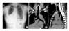
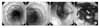
The chest x-ray revealed atelectasis of the right middle and lower lobes of the lung (Figure 1). The chest computerized tomography (CT) showed no demonstrable active parenchymal lesion in both lungs with no enlarged bronchial artery. Therefore we could not try bronchial artery embolization. A bronchoscopy was done and disclosed a very large blood clot totally obstructing the right bronchus intermedius (Figure 2). Incidentally, abnormal nodular projections of the tracheal anterolateral wall from just below vocal cord to 2 cm above carina were identified and tracheal biopsy was performed. After the verification of no other endobronchial abnormalities or an active bleeding focus, bronchoscopic removal of the blood clot was performed. The biopsy findings were chronic inflammation with a mild dystrophic calcification and a moderate degree of squamous cell dysplasia. The symptoms and atelectasis improved and the patient was discharged 10 days after admission.
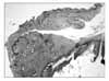
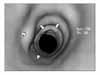
Three months later, the patient returned for follow up. The chest CT showed no definite consolidation or mass in the lungs. The bronchoscopy showed no significant interval change of the multiple nodules in the tracheal wall. A second biopsy specimen was obtained. The pathologic findings were consistent with a submucosal osteochondromatous nodule compatible with TO (Figure 3). The diagnosis of TO was made and the patient continued to be free of symptoms. Thirty-two months later, the patient had a second attack of massive hemoptysis with atelectasis at the same sites. The virtual bronchoscopy showed multiple tracheal nodules and near total obstruction of the bronchus intermedius (Figure 4). The third bronchoscopic examination showed no interval change of the TO and the blood clot was removed successfully. The patient reported taking an aspirin 100 mg once daily for the last one year without prescription and this was discontinued. The patient is followed closely and is currently doing well.
Discussion
Tracheopathia osteochondroplastica (TO) is a rare disorder of the trachea and large airways of unknown etiology; it is frequently found as an incidental autopsy finding. Recently, clinical diagnosis has been more common due to the frequent use of fiberoptic bronchoscopy and chest CT. The incidence of TO at autopsy was estimated of approximately 3/1000, while data from bronchoscopy were widely varying from 1/125 to 1/60005. Most affected patients have non-specific respiratory symptoms such as cough, hemoptysis, dyspnea, wheezing, or recurrent infection2,3. Sometimes patients with TO are identified after difficulty with intubation because of a narrowed trachea6 or a bronchoscopy done for other reasons. In Korea, eleven cases of TO were reported until now3,7-12. Most of the patients had mild non-specific symptoms such as chronic cough, dyspnea, hoarseness or respiratory infection. We could not find the TO case with atelectasis due to massive hemoptysis.
The diagnosis of a TO can be made by fiberoptic bronchoscopy; the findings of this disorder are unique and include multiple osteocartilaginous calcified nodules within the submucosa of the anterior and lateral aspects of the tracheobronchial tree1,2; the membranous portions of the trachea are characteristically spared in contrast to tracheobronchial amyloidosis13. Yokoyama et al described the multiple osteocartilaginous nodules as "rock garden" findings14. Chest CT, magnetic resonance imaging (MRI) or virtual bronchoscopy can all detect these submucosal nodules in the trachea and main bronchi; in half of the cases the nodules are calcified.
A pathological diagnosis is desirable, but not necessary for confirmation of TO. Biopsy specimens can be difficult to obtain by flexible bronchoscopy due to the hard consistency of the lesions14. The histopathology of these nodules shows calcifications, chondrifications or ossifications of the upper layer of the mucous membranes. The cartilage of the TO nodules can be distinguished from normal cartilaginous rings; the bone tissues may show bone marrow foci with active areas of hematopoiesis15. In half of the cases, squamous metaplasia of the tracheal epithelium is found2.
Although rapid progression of TO has been reported16, most cases progress very slowly2. There is no specific treatment for prevention of nodular formation or progression available to date. The majority of patients do well with conservative management such as antibiotic treatment, mucolytics or medical hemostasis. A recent report reviewed many therapeutic interventions such laser resection, endoscopic removal of the nodules, radiation therapy, and surgical repair or placement of stents4,15.
 XML Download