

 PDF
PDF ePub
ePub Citation
 Print
Print


Dong Won Shin, M.D.1, Moon Han Choi, M.D.1, Seung Sik Park, M.D.1, Sung Woo Park, M.D.1, Ki Up Kim, M.D.1, An Soo Jang, M.D.1, Choon-Sik Park, M.D.1 , Cheol Wan Lim, M.D.2, Eun Suk Ko, M.D.3, Sang Hyun Paik, M.D.4, Do Jin Kim, M.D.1
, Cheol Wan Lim, M.D.2, Eun Suk Ko, M.D.3, Sang Hyun Paik, M.D.4, Do Jin Kim, M.D.1
Abstract
The incidence of appendiceal metastatic cancer is quite low. In particular, in small cell lung cancer, there is a very low incidence of a metastasis to the appendix. A 75-years old man with right lower quadrant pain, cough and sputum was transferred to our hospital. Abdominal CT revealed acute appendicitis with a perforation. The patient underwent surgery. The frozen sections of the tissue obtained during surgery, indicated a malignancy, but a right hemicolectomy was not performed due to the patient's poor general condition. The histology findings of the appendix were identified as a small cell carcinoma. The abdominal CT scan and chest x-ray at admission day showed a mass in the right lower lobe, and a further evaluation of the lesion was performed including positron emission tomography and flexible bronchoscopy with a biopsy. The pathology findings of the lung mass were also small cell lung cancer. The specimens from both sites stained positive for cytokeratin, cluster designation 56, synaptophysin, chromogranin-A and thyroid transcription factor 1. It was concluded that the appendiceal small cell cancer originated from the lung.
Figures and Tables
Figure 1
Abdominal enhanced CT finding shows a distended appendix (1.4 cm) with wall thickening and fat infiltration around appendix.
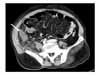
Figure 2
In follow-up chest CT after 8 days showed about 4.5 cm sized mass with ill-defined margin and increased pleural effusion.
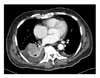
Figure 3
Post-operative PET-CT scan reveals hypermetabolic activity in right lower paratracheal lymph node, right lower lobe, right adrenal gland and right anterior abdominal wall (clockwise direction from upper left panel). Beside abdominal wall, other sites were considered as malignancy with the calculated SUVs more than 3.6.
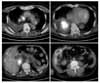
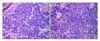
Figure 4
Microscopic findings. (A) Appendix. Light microscopic findings shows round shaped small cells with dense nuclei, inconspicuous nucleoli, and sparse cytoplasm (H&E stain, ×400). (B) Light microscopic finding of bronchoscopic biopsy tissue shows similar features as appendix (H&E stain, ×200).

References
1. Cohen MH, Matthews MJ. Small cell bronchogenic carcinoma: a distinct clinicopathologic entity. Semin Oncol. 1978. 5:234–243.
2. Sridhar KS, Hussein AM, Thurer RJ. Evolving role of surgical treatment in limited-disease small cell lung carcinoma. J Surg Oncol. 1989. 40:155–161.
3. Johnson BE. Management of small cell lung cancer. Clin Chest Med. 2002. 23:225–239.
4. Pang LC. Metastasis-induced acute appendicitis in small cell bronchogenic carcinoma. South Med J. 1988. 81:1461–1462.
5. Goldstein EB, Savel RH, Walter KL, Rankin LF, Satheesan R, Lehman HE, et al. Extensive stage small cell lung cancer presenting as an acute perforated appendix: case report and review of the literature. Am Surg. 2004. 70:706–709.
6. Levchenko AM, Vasechko VN, Erusalimskiĭ EL. Metastasis of small-cell lung cancer to the appendix. Klin Khir. 1985. 5:56–57.
7. Park IJ, Yu CS, Kim HC, Kim JC. Clinical features and prognostic factors in primary adenocarcinoma of the appendix. Korean J Gastroenterol. 2004. 43:29–34.
8. Cortina R, McCormick J, Kolm P, Perry RR. Management and prognosis of adenocarcinoma of the appendix. Dis Colon Rectum. 1995. 38:848–852.
9. Ryo H, Sakai H, Ikeda T, Hibino S, Goto I, Yoneda S, et al. Gastrointestinal metastasis from lung cancer. Nihon Kyobu Shikkan Gakkai Zasshi. 1996. 34:968–972.
10. Lobins R, Floyd J. Small cell carcinoma of unknown primary. Semin Oncol. 2007. 34:39–42.
11. Lau SK, Luthringer DJ, Eisen RN. Thyroid transcription factor-1: a review. Appl Immunohistochem Mol Morphol. 2002. 10:97–102.
12. Ordonez NG. Value of thyroid transcription factor-1 immunostaining in distinguishing small cell lung carcinomas from other small cell carcinomas. Am J Surg Pathol. 2000. 24:1217–1223.
13. Fletcher MS. Gastric perforation secondary to metastatic carcinoma of the lung: a case report. Cancer. 1980. 46:1879–1882.
14. Morgan MW, Sigel B, Wolcott MW. Perforation of a metastatic carcinoma of the jejunum after cancer chemotherapy. Surgery. 1961. 49:687–689.
15. Shen YY, Shiau YC, Wang JJ, Ho ST, Kao CH. Whole-body 18F-2-deoxyglucose positron emission tomography in primary staging small cell lung cancer. Anticancer Res. 2002. 22:1257–1264.
 XML Download