

 PDF
PDF ePub
ePub Citation
 Print
Print


Abstract
Background
Congenital cystic adenomatoid malformation of the lung (CCAM) is a rare congenital developmental anomaly of the lower respiratory tract. Most cases are diagnosed within the first 2 years of life, so adult presentation of CCAM is rare. We describe here six adult cases of CCAM and the patients underwent surgical resection, and all these patients were seen during a five and a half year period. The purpose of this study was to analyze the clinical, radiological and histological characteristics of adult patients with CCAM.
Methods
Through medical records analysis, we retrospectively reviewed the clinical characteristics, the chest pictures (X-ray and CT) and the histological characteristics.
Results
Four patients were women and the mean age at diagnosis was 23.5 years (range: 18~39 years). The major clinical presentations were lower respiratory tract infection, hemoptysis and pneumothorax. According to the chest CT scan, 5 patients had multiseptated cystic lesions with air fluid levels and one patient had multiple cavitary lesions with air fluid levels, and these lesions were surrounded by poorly defined opacities at the right upper lobe. All the patients were treated with surgical resection. 5 patients underwent open lobectomy and one patient underwent VATS lobectomy. On the pathological examination, 3 were found to be CCAM type I and 3 patients were CCAM type II, according to Stocker's classification. There was no associated malignancy on the histological studies of the surgical specimens.
Conclusion
As CCAM can cause various respiratory complications and malignant changes, and the risks associated with surgery are extremely low, those patients who are suspected of having or who are diagnosed with CCAM should go through surgical treatment for making the correct diagnosis and administering appropriate treatment.
Figures and Tables
Figure 1
There are multiple cystic lesions, about 6~7 cm in diameter in the left middle lung field. Pneumonic consolidations and air fluid levels are in and around the cystic lesions (A). At the level of the left inferior pulmonary vein, the lesion contains about 4.5 cm in diameter cystic space (B). The CT scan at the level of left main bronchus reveals innumerable fine cystic lesions in the superior segment of the left lower lobe (C).
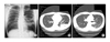
Figure 2
Chest PA shows a cystic lesion with air-fluid level and pericystic pneumonic infiltration in right lower lobe on admission (A). Chest CT scan on admission reveals a thin walled multicystic lesion with fluid collection mimics a pulmonary abscess in RLL (B).
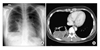
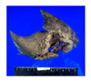
Figure 3
Gross finding of a resected right lower lobe in Case 6 shows one large cystic lesion and small variable sized cysts surrounding by bronchial like structures, accompanied with grayish pneumonic consolidation.

References
1. Stocker JT, Madewell JE, Drake RM. Congenital cystic adenomatoid malformation of the lung: classification and morphologic spectrum. Hum Pathol. 1977. 8:155–171.
2. Rosado-de-Christenson ML, Stocker JT. Congenital cystic adenomatoid malformation. Radiographics. 1991. 11:865–886.
3. Luján M, Bosque M, Mirapeix RM, Marco MT, Asensio O, Domingo C. Late-onset congenital cystic adenomatoid malformation of the lung: embryology, clinical symptomatology, diagnostic procedures, therapeutic approach and clinical follow-up. Respiration. 2002. 69:148–154.
4. Laberge JM, Flageole H, Pugash D, Khalife S, Blair G, Filiatrault D, et al. Outcome of the prenatally diagnosed congenital cystic adenomatoid lung malformation: a Canadian experience. Fetal Diagn Ther. 2001. 16:178–186.
5. d'Agostino S, Bonoldi E, Dante S, Meli S, Cappellari F, Musi L. Embryonal rhabdomyosarcoma of the lung arising in cystic adenomatoid malformation: case report and review of the literature. J Pediatr Surg. 1997. 32:1381–1383.
6. Federici S, Domenichelli V, Tani G, Sciutti R, Burnelli R, Zanetti G, et al. Pleuropulmonary blastoma in congenital cystic adenomatoid malformation: report of a case. Eur J Pediatr Surg. 2001. 11:196–199.
7. Kaslovsky RA, Purdy S, Dangman BC, McKenna BJ, Brien T, Ilves R. Bronchioloalveolar carcinoma in a child with congenital cystic adenomatoid malformation. Chest. 1997. 112:548–551.
8. Ribet ME, Copin MC, Soots JG, Gosselin BH. Bronchioloalveolar carcinoma and congenital cystic adenomatoid malformation. Ann Thorac Surg. 1995. 60:1126–1128.
9. Oh BJ, Lee JS, Kim JS, Lim CM, Koh Y. Congenital cystic adenomatoid malformation of the lung in adults: clinical and CT evaluation of seven patients. Respirology. 2006. 11:496–501.
10. Ch'in KY, Tang MY. Congenital adenomatoid malformation of one lobe of a lung with general anasarca. Arch Pathol (Chic). 1949. 48:221–229.
11. Morelli L, Piscioli I, Licci S, Donato S, Catalucci A, Del Nonno F. Pulmonary congenital cystic adenomatoid malformation, type I, presenting as a single cyst of the middle lobe in an adult: case report. Diagn Pathol. 2007. 2:17.
12. MacSweeney F, Papagiannopoulos K, Goldstraw P, Sheppard MN, Corrin B, Nicholson AG. An assessment of the expanded classification of congenital cystic adenomatoid malformations and their relationship to malignant transformation. Am J Surg Pathol. 2003. 27:1139–1146.
13. Lantuejoul S, Ferretti GR, Goldstraw P, Hansell DM, Brambilla E, Nicholson AG. Metastases from bronchioloalveolar carcinomas associated with longstanding type 1 congenital cystic adenomatoid malformations: a report of two cases. Histopathology. 2006. 48:204–206.
14. Weatherford DA, Stephenson JE, Taylor SM, Blackhurst D. Thoracoscopy versus thoracotomy: indications and advantages. Am Surg. 1995. 61:83–86.
15. Kwon YS, Koh WJ, Han J, Choi YS, Kim K, Kim J, et al. Clinical characteristics and feasibility of thoracoscopic approach for congenital cystic adenomatoid malformation in adults. Eur J Cardiothorac Surg. 2007. 31:797–801.
16. Allan BT, Day DL, Dehner LP. Primary pulmonary rhabdomyosarcoma of the lung in children: report of two cases presenting with spontaneous pneumothorax. Cancer. 1987. 59:1005–1011.
17. Granata C, Gambini C, Balducci T, Toma P, Michelazzi A, Conte M, et al. Bronchioalveolar carcinoma arising in congenital cystic adenomatoid malformation in a child: a case report and review of malignancies originating in congenital cystic adenomatoid malformation. Pediatr Pulmonol. 1998. 25:62–66.
 XML Download