

 PDF
PDF ePub
ePub Citation
 Print
Print


Introduction
Bronchial foreign body aspiration usually occurs in children younger than 3 years old or adults with weakened tracheal defense mechanism but it also rarely occurs in healthy adults. Acute complications of endobronchial foreign body aspiration include asphyxia, cardiac arrest, and pneumothorax while chronic complications range from pneumonia, lung abscess, bronchiectasis and hemoptysis to bronchial stenosis. And thus, early diagnosis and treatment are critical in preventing such complications. However, if the aspiration of a radiolucent foreign body has occurred in a healthy adult patient and has not been recognized by the patient, the diagnosis process becomes a challenging work.
Case Report
A 62-year-old male with tobacco abuse (66 pack-years) was presented to our hospital with symptoms of cough, blood tinged sputum and dyspnea. On physical examination, breathing sounds were coarse and wheezes were heard on right side in the lower lung fields. On peripheral blood examination, the white blood cell count was 6,900/mm3 (neutrophil 47%) while the hemoglobin and the platelet count was 15.5 g/dl and 274,000/mm3 respectively. The arterial blood gas levels were pH 7.43, PCO2 42 mmHg, PaO2 72 mmHg, HCO3- 26 mmol/L and SaO2 96%. On pulmonary function test, FVC was 2.49 L (71% predicted), FEV1 1.64 L (68% predicted) and FEV1/FVC 68%.
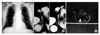
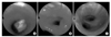
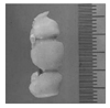
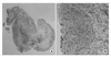
Initial chest x-ray had shown no suspicious evidence of foreign body aspiration (Figure 1A). Chest CT was presented with a focal wall thickening and linear high density in the lumen of right bronchus intermedius (Figure 1B). Additionally, a flexible bronchoscopic examination was conducted to identify the endobronchial lesion and it revealed a foreign material in the proximal portion of right bronchus intermedius (Figure 2A). It was removed using rat-tooth forceps and was revealed as a temporary resin bridge which is often used as an orthodontics tool (Figure 3). Then a biopsy was performed since multiple polypoid protruding masses were observed distal to the impacted site (Figure 2B) and on pathology, localized dysplastic change, granuloma formation and inflammatory cell infiltration were observed (Figure 4).
History taking was done once again in an effort to track the process of the actual aspiration via interview with the patient and he provided an affirmation that he did lose his temporary resin bridge while he was coughing 2 months ago.
Subsequently, we performed a separate CT on the bronchial foreign body removed by bronchoscopy and this CT showed high density in the foreign material (Figure 1C), which was radiolucent in the previous chest x-ray. This finding enabled us to form a correlation between the result from this CT and the linear high density in the lumen of right bronchus intermedius in the previous chest CT (Figure 1B). Two months later, we conducted the same bronchoscopic examination and at this time, only mild mucosal elevation was found, without any of the previously noted masses being revealed (Figure 2C).
Discussion
In dealing with the case described above, we had challenges in identifying the presence of foreign body in the patient's bronchus. A variety of foreign materials such as bone fragments, teeth, dentures, pins, vegetable matter, food particles, and nuts can be aspirated in some cases, but this rarely happens with adults, especially when they are in a conscious and awaken state. The patient in this case did not have a history of depressed sensorium or loss of consciousness and what made the discovery process even more difficult was that the event of aspiration was obscure. This primarily caused challenges in leading to the suspicion of potential foreign body aspiration. The second misleading factor was that the results of the chest x-ray were normal. Typically, while just a simple chest x-ray can lead to discovery of radiolucent foreign body aspiration such as an obstructive emphysema, atelectasis, bronchiectasis, localized pneumonia and hyperinflation during inspiration, it is more common to get normal findings in chest x-rays1. And this is supported by Sersar et al2, who reported 3,300 patients with bronchial foreign body aspiration, and out of these only 23.5% actually revealed the presence of foreign materials via a simple chest x-ray. CT is a more sensitive tool in diagnosing radiolucent foreign body aspiration than simple x-ray. Applegate3 reported that after aspirating LEGO (a plastic block), one of the radiolucent materials, CT revealed a sensitivity of 83% and a specificity of 89%. Otherwise food material, such as peanuts showed a lower sensitivity of 34% and a specificity of 89%.
In some cases, however, both simple x-ray and CT failed to provide any findings of radiolucent materials, thereby making the discovery process very difficult. Therefore, it is crucial to keep in mind the possibility of potential radiolucent foreign body aspiration even when the radiographic finding is normal if a patient has recurrent fever, cough, sputum, blood tinged sputum and chest pain but does not respond to treatments and rather shows complications of an unknown origin. In such situations, the use of an alternative diagnostic tool such as bronchoscopy should be considered as an option.
Currently used prosthetic resins are radiolucent and thus are difficult to be captured in an image form with standard radiographic techniques. Recently, radiopaque dental additive materials, such as triphenylbismuth, were developed to overcome this problem4.
In the case of our patient, interestingly, inflammatory polyps were found on the site impacted by entry of the foreign material. Inflammatory polyps, which are a nontumorous lesion, develop due to fibrotic tissue proliferation and usually occur in association with endobronchial stimulants and hot gas or corrosive material inspiration5. Greene et al6 reported that aspiration of sunflower seeds causes inflammatory polyps and Berman et al7 reported that a plastic piece aspiration leads to the same result as with sunflower seeds. Although focal dysplastic changes were noted by microsopic examination, spontaneous regression occurred after removal of the foreign material. Eventually, the polyps may be the consequence of a hyper-regenerative process following mucosal irritation by resin compounds.
 XML Download