Abstract
The patient is a 62-year-old man with known diabetes mellitus who presented with a two-weeks-history of dyspnea, cough, and fever. He was diagnosed with a lung abscess in the right upper lobe and was treated with intravenous antibiotics. The patient's clinical and radiological findings improved within seven days after medical treatment. However, newly developed ground-glass opacity and infiltrations were observed in the right lower lung. Fourteen days after admission, the patient's symptoms and imaging finding became aggravated despite trestment with susceptible antibiotics for lung abscess. Trans-bronchial lung biopsy (TBLB) was performed in the lateral basal segment of the right lower lobe of the lung. A histologic photomicrograph showed organizing pneumonia, also called bronchiolitis obliterans with organizing pneumonia(BOOP), that became more definite as the terminal bronchioles and alveoli became occluded with masses of inflammatory cells and fibrotic tissue. The clinical symptoms and radiograph findings resolved quickly with prednisone treatment. We report a case of secondary organizing pneumonia diagnosed after TBLB following lung abscess treatment and provide a review of the literature.
Figures and Tables
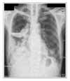
 | Figure 1Chest radiograph and computed tomography showing a loculated lung absecss in the right upper lobe. |
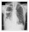
 | Figure 2Chest radiograph and CT scan showing air-fluid level characteristic of lung abscess and septated lesion in the right upper lobe. Clinical and imaging improvement was obtained with intravenous antibiotics treatment. |
References
1. Kim CH, Cha SI, Han CD, Kim YJ, Lee YS, Park JY, et al. Percutaneous catheter drainage of lung abscess. Tuberc Respir Dis. 1993. 40:158–164.
2. Hagan JL, Hardy JD. Lung abscess revisited. A survey of 184 cases. Ann Surg. 1983. 197:755–762.
3. Epler GR, Colby TV, McLoud TC, Carrington CB, Gaensler EA. Bronchiolitis obliterans organizing pneumonia. N Engl J Med. 1985. 312:152–158.
4. Chang JH, Park SY. Twenty four cases of idiopathic bronchiolitis obliterans organizing pneumonia, reported in Korea and a review of literatures. Tuberc Respir Dis. 1999. 46:709–717.
5. American Thoracic Society and European Respiratory Society. American Thoracic Society/European Respiratory Society International Multidisciplinary Consensus Classification of the Idiopathic Interstitial Pneumonias. Am J Respir Crit Care Med. 2002. 165:277–304.
6. Cordier JF. Cryptogenic organising pneumonia. Eur Respir J. 2006. 28:422–446.
7. Cohen AJ, King TE, Downey GP. Rapidly progressive bronchiolitis obliterans with organizing pneumonia. Am J Respir Crit Care Med. 1994. 149:1670–1675.
8. Cordier JF. Organizing pneumonia. Thorax. 2000. 55:318–328.
9. Epler GR. Bronchiolitis obliterans organizing pneumonia. Semin Respir Infect. 1995. 10:65–77.
10. Lee JH, Park MJ, Kim YH, Park BJ, Oh WT, Lee MY, et al. Two cases of bronchiolitis obliterans organizing pneumonia treated with steroid and cyclosporine therapy. Tuberc Respir Dis. 2005. 59:315–320.
11. Miyagawa-Hayashino A, Wain JC, Mark EJ. Lung transplantation biopsy specimens with bronchiolitis obliterans or bronchiolitis obliterans organizing pneumonia due to aspiration. Arch Pathol Lab Med. 2005. 129:223–226.


 PDF
PDF ePub
ePub Citation
 Print
Print


 XML Download