

 PDF
PDF ePub
ePub Citation
 Print
Print


INTRODUCTION
The recent increase in tuberculosis cases and the emergence of multi-drug resistant strains have called for more rapid and sensitive methods in the laboratory diagnosis than the conventional diagnostic techniques1-3. Although the culture of Mycobacterium tuberculosis (MTB) continues to be the gold standard for the diagnosis of tuberculosis, the results are neither satisfactorily sensitive nor rapid. The mycobacterial culture takes at least two weeks or longer depending on culture media used4. The nucleic acid amplification tests (NATs) including polymerase chain reaction (PCR) improved the accuracy as well as the time for the diagnosis of tuberculosis in respiratory specimens. For nonrespiratory specimens like pleural effusion, however, previous studies have shown highly variable results about the usefulness of NAT because they used different in-house NAT methods, rather small study populations, and diverse criteria of pleural tuberculosis.
The Cobas Amplicor MTB test (Roche Diagnostic Systems, Inc., Branchburg, NJ, USA) is a well-established and commercially available PCR technique commonly used for the direct detection of M. tuberculosis in clinical samples. The test uses biotinylated genus-specific primers (KY18 and KY75) to amplify the 584-base-pair sequence within the 1500-base-pair region encoding 16s rRNA of M. tuberculosis. It combines five instruments into one (thermal cycler, automatic pipettor, incubator, washer and reader). Because it is fully automated for the amplification and detection steps on a single instrument, it is thought to be able to minimize inter-individual variabilities. Furthermore, since it binds M. tuberculosis-specific oligonucleotide probes to the amplified sequences, it can increase the overall specificity.
Tuberculous pleural effusion occurs in up to 30% of patients with tuberculosis5 and occupies the major portion of the extrapulmonary tuberculosis morbidity6. However, the number of organisms in the pleural effusion from most cases of tuberculous pleuritis is relatively low, with positive cultures found in less than 25% of cases. Even the pleural biopsy shows granulomatous inflammation only in approximately 60% of patients7. PCR has been used to detect M. tuberculosis in pleural fluid samples, with highly variable sensitivities (11 to 81%) in the previous studies using different in-house PCR methods8-13. Our previous study has, however, demonstrated very low sensitivity of PCR technique for the diagnosis of pleural tuberculosis (sensitivity 17.5%, specificity 98.1%) using the kit developed for the diagnosis of pulmonary tuberculosis14. The purpose of this investigation is to determine whether the sensitivity of PCR technique for the diagnosis of pleural tuberculosis can be improved when increasing the amount of pleural effusion specimens.
MATERIALS AND METHODS
Study population and samples
Fifty three patients older than 18 years of age, for whom the exclusion of the possibility of tuberculous pleural effusion was necessary, were prospectively analyzed for one year. The suspicion of pleural tuberculosis was based on the unilateral pleural effusion with clinical manifestations suggestive of tuberculosis such as productive cough, chronic low-grade fever, weight loss, anorexia, night sweating, etc. All of the 53 pleural effusion specimens were sent for routine analysis, acid-fast bacilli (AFB) smear, mycobacterial culture, adenosine deaminase (ADA) level, Gram stain, bacterial culture, and cytologic examination. For the comparison of the sensitivities of pleural effusion PCR test according to the amount of pleural effusion specimens, M. tuberculosis PCR was performed with 10, 25, and 50 ㎖ of each pleural effusion sample, respectively. The sputum was collected in the morning from each patient and was sent for AFB smear and mycobacterial culture. Pleural biopsy and pathologic examination were not performed in 36 out of the 53 patients due to a small amount of pleural effusion and/or patient's refusal.
Diagnostic criteria of pleural tuberculosis
Cases of pleural tuberculosis were defined as those with one of the following: positive M. tuberculosis culture of pleural fluid, and/or histopathologic finding consistent with tuberculosis on pleural biopsy, and/or positive M. tuberculosis culture of sputum, and/or positive M. tuberculosis culture of other biologic specimens, and/or positive response to anti-tuberculous medication without other possible causes of pleural effusion.
AFB smear and mycobacterial culture
For a microscopic examination, Ziehl-Neelsen staining was performed. After being decontaminated by an equal volume of 4% sodium hydroxide (NaOH) solution, each of the collected sputum and pleural effusion samples was inoculated onto two slopes of Ogawa media containing 3% potassium dihydrogen phosphate (KH2PO4) (3% Ogawa media)15. The inoculated medium was incubated at 35-37℃ until the growth was observed or was discarded as negative after eight weeks.
Determination of adenosine deaminase (ADA) activity in pleural effusion
ADA activity was determined using 2 ml of pleural fluid by the colorimetric method described by Giusti16. ADA level below 45 IU/L was considered as negative.
Tuberculin skin test
Tuberculin skin test was not included in this study because there were difficulties in interpreting the results in Korea where Mycobacterium bovis BCG vaccination program covers more than 90% of the population.
Nucleic acid amplification and detection techniques
(i) Specimen preparation: pleural fluid specimens were decontaminated using the equal amount of 4% NaOH solution and were centrifuged at 3,000 x g for 20 minutes to collect the sediments. 100 ㎕ of sediment from each pleural effusion sample was transferred to a microcentrifuge tube containing 500 ㎕ of washing solution, and centrifuged at 12,500 × g for 10 minutes. The supernatant was discarded and 100 ㎕ of specimen lysis reagent was added to extract DNA template. The mixture was vortexed and incubated at 60℃ in a dry heat block for 45 minutes. 100 ㎕ of specimen neutralization reagent was added. Then a 50-㎕ aliquot of the DNA extract was transferred to a PCR tube containing 50 ㎕ of amplification mixture.
(ii) Amplification, hybridization, detection, and interpretation were performed according to the manufacturer's instructions.
RESULTS
Subjects' characteristics
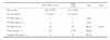
Of the 53 patients, 26 (49.1%) were diagnosed with pleural tuberculosis. The cases of pleural tuberculosis (n=26) consisted of those with positive M. tuberculosis culture of pleural fluid (n=4), those with histopathologic finding consistent with tuberculosis on pleural biopsy (n=11), those with positive M. tuberculosis culture of sputum (n=6), and/or those with a positive response to anti-tuberculous medication (n=12) without other possible causes of pleural effusion. The mean age was 44.2 years for the pleural tuberculosis patients and 60.2 years for the non-tuberculous pleural effusion patients. The number of female patients was 12 (46.2%) and 8 (29.6%) in each group (Table 1). The etiologies of the non-tuberculous pleural effusion included malignancy (n=10, 37.0%), bacterial pneumonia (n=11, 40.8%), sepsis (n=2, 7.4%), intra-abdominal infection (n=1, 3.7%), and undetermined origin (n=3, 11.1%).
Sensitivity and specificity of pleural effusion AFB smear, MTB culture, ADA activity, pleural biopsy pathology, and sputum MTB culture
Of the 26 tuberculous pleural effusion specimens, AFB smear-positive was one, MTB culture-positive were four, and ADA-positive (above 45 IU/L) were 25. Considering the combination of MTB culture, pleural pathology and clinical diagnosis as the reference method of diagnosing pleural tuberculosis, the sensitivities were 3.8%, 15.4%, and 96.2%, respectively. The pleural biopsy was performed in 13 of the 26 pleural tuberculosis patients, and 11 of the 13 pleural biopsy-performed patients showed the histologic findings consistent with pleural tuberculosis. Of the 26 pleural tuberculosis patients, six showed the sputum MTB culture-positive findings. The sensitivities of pleural biopsy and sputum MTB culture were 84.6% and 23.1%, respectively. Of the 27 non-tuberculous pleural effusion specimens, ADA-positive were eight. The specificity of pleural effusion ADA activity, therefore, was 70.4% (Table 1).
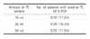
Sensitivities of pleural effusion MTB PCR test according to the amount of pleural effusion specimens
Of the 26 tuberculous pleural effusion specimens, MTB PCR-positive were three, four, and three when using 10 ml, 25 ml, and 50 ml of pleural fluid samples, respectively. The sensitivities of pleural effusion MTB PCR test were 11.5%, 15.4%, and 11.5%, respectively, which did not show statistically significant differences (p>0.05, symmetry exact test) (Table 2).
Of the four pleural effusion MTB PCR-positive patients, pleural effusion AFB smear-positive was one, pleural effusion MTB culture-positive were three, pleural biopsy pathology-positive were three (pleural biopsy was not performed to the remaining one), and sputum MTB culture-positive were two.
DISCUSSION
As for tuberculous pleurisy, early in the course of tuberculous infection, a few organisms may gain access to the pleural space and cause a hypersensitivity response in the presence of cell-mediated immunity17,18. Commonly, this form of tuberculous pleuritis goes unnoticed, and it resolves spontaneously. In some patients, however, the tuberculous involvement of pleura is manifested as an acute illness with fever and pleuritic pain. The effusion is generally small and unilateral. In approximately 30% of patients, there is no radiographic evidence of involvement of the lung parenchyma in spite of the presence of lung parenchymal lesions in most cases as evidenced by findings of lung dissections19. In the absence of concurrent pulmonary tuberculosis, the diagnosis of pleural tuberculosis requires thoracentesis and, in most cases, even pleural biopsy7.
For tuberculous pleural effusion, the number of organisms in the pleural fluid is very small, so the conventional methods for the detection of M. tuberculosis are often of no use. Even though the combination of microscopic examination and culture of pleural biopsy specimens was reported to increase the diagnostic rate up to 90%, it is time-consuming20. Thus many physicians request nucleic acid amplification tests (NATs) including PCR for pleural effusion specimens to obtain a rapid and accurate diagnosis of pleural tuberculosis.
Several commercial and in-house NATs to detect MTB in clinical specimens, have been developed. These tests amplify various targets in DNA or RNA sequences that are genus- or species-specific, which is followed by the detection step using gel electrophoresis or hybridization method. Currently there are four commercial NATs for the detection of MTB: 1) the Amplicor MTB test and its automated version, the Cobas Amplicor MTB test (Roche Diagnostic Systems, Inc., Branchburg, NJ), 2) the Enhanced Amplified Mycobacterium Tuberculosis Direct Test (E-AMTDT) (Gen-Probe, Inc., San Diego, CA), 3) the BDProbe Tec ET test (Becton Dickinson, Sparks, MD), and 4) the INNO-LiPA-Rif. TB test (Innogenetics N. V., Zwijndrecht, Belgium). Of these, the E-AMTDT was approved by the FDA for the direct detection of MTB in both smear-positive and smear-negative respiratory specimens from the patients suspected of having tuberculosis, and the Amplicor MTB test was approved only for smear-positive respiratory specimens21-23. As for in-house NATs, diverse methods using different primers have been used to detect MTB in pleural fluid samples, with highly variable sensitivities (11 to 81%) in the previous studies8-13.
As for commercial NATs, the sensitivities have also been variable (20 to 100%) because the number of the patients with pleural tuberculosis was rather small and, in some studies, only the MTB culture-positive cases were included in the pleural tuberculosis group24-30. Because the cases with MTB culture-positive pleural effusion occupy a relatively small portion of the whole pleural tuberculosis cases and the sensitivity of the pleural effusion MTB PCR test largely depends on the bacillary load, the sensitivity from the study group of patients with MTB culture-positive pleural effusion does not reflect the exact one from the whole group of patients with pleural tuberculosis. From these reasons, there has been no consensus made about the usefulness of the MTB PCR test in the diagnosis of tuberculous pleural effusions.
In our previous study, we have demonstrated the low sensitivity of MTB PCR test for the diagnosis of pleural tuberculosis (sensitivity 17.5%, specificity 98.1%), using the commercially available Cobas Amplicor MTB test which can minimize the inter-tester variabilities and maximize the specificities due to the automated specimen processing and detection step and employing the combination of MTB culture, pleural pathology and clinical diagnosis as the reference method of diagnosing pleural tuberculosis14.
In this study, we have examined whether the sensitivity of pleural effusion MTB PCR test for the diagnosis of pleural tuberculosis can be improved when increasing the amount of pleural effusion specimens. The results again showed that MTB PCR test of pleural effusion has a lower sensitivity compared with mycobacterial culture of pleural effusion sample and pleural biopsy pathology. The sensitivities of pleural effusion MTB PCR test using a different amount of pleural fluid samples, did not show statistically significant differences. These results indicate that the sensitivity of MTB PCR test cannot be improved even with an increased amount of pleural effusion specimens. The sensitivity of NAT depends not only on the number of mycobacteria but also on their homogenous distribution in the specimen, the presence of the amplification inhibitor in the sample, and the type of the primers31. When applying MTB PCR test to paucibacillary specimens, therefore, these all aspects should also be considered.
Although MTB PCR assay provides a rapid result and has a potential role in confirming tuberculous pleuritis, in conclusion, it has limitations in itself. Our results suggest that the pleural effusion MTB PCR using the Cobas Amplicor MTB test has a low sensitivity and hence does not seem to be useful in excluding the disease. Therefore, it cannot replace the conventional diagnostic methods including culture techniques and histopathologic examinations. Furthermore, the results of the pleural effusion MTB PCR test need to be interpreted in conjunction with those of the conventional methods and the clinical findings to reach the final diagnosis of pleural tuberculosis.
 XML Download