

 PDF
PDF ePub
ePub Citation
 Print
Print


Abstract
Endobronchial foreign bodies are difficult to diagnose as the cause of obstructive pneumonia and atelectasis, However, once discovered, they can generally be removed, leading to an immediate and dramatic resolution of the symptoms. Occasionally, small foreign bodies that lodge in the peripheral airway are often initially asymptomatic but become symptomatic several years later.
We reported a case of obstructive pneumonia and massive hemoptysis caused by a foreign metallic body. The patient knew that the foreign body was lodged in the peripheral airway on the chest X-ray, but did not want treatment. Several years later, he had a massive hemoptysis and obstructive pneumonia. Removal with a flexible bronchoscope failed, but the metallic foreign body was self-expectorated by coughing after the procedure. The pneumonia was resolved after removing the foreign body. The patient improved and was discharged without any sequela.
Go to : 
REFERENCES
1.Son CY., Wee JO., Kim SO., Oh IJ., Park CM., Kim KS, et al. A retrospective review of tracheobronchial foreign bodies. Tuberc Respir Dis. 2005. 58:600–6.

2.Jo KG., Baek MS., Kim MS., Hur JM., Jeon JI., Park KS, et al. A case of occult foreign body lodged in bronchus for a long period and removal by flexible bronchoscopy. Tuberc Respir Dis. 1997. 44:1166–71.
3.Lan RS. Non-asphyxiating tracheobronchial foreign bodies in adults. Eur respire J. 1994. 7:510–4.
4.Chen CH., Lai CL., Tsai TT., Lee YC., Perng RP. Foreign body aspiration into the lower airway in Chinese adults. Chest. 1997. 112:129–33.
5.al Majed SA., Ashour M., al Mobeireek AF., al Hajjaj MS., Alzeer AH., al Kattan K. Overlooked inhaled foreign bodies: late sequelae and the likelihood of recovery. Respir Med. 1997. 91:293–6.
6.Baharloo F., Veyckemans F., Francis C., Biettlot MP., Rodenstein DO. Tracheobronchial bodies: presentation and management in children and adults. Chest. 1999. 115:1357–62.
7.Limper AH., Prakash UB. Tracheobronchial foreign bodies in adults. Ann Intern Med. 1990. 112:604–9.
8.Lee BJ., Lee YW., Jung JW., Shin JW., Kim JY., Park IW, et al. A case of bronchial foreign body misdiagnosed as bronchial asthma. Tuberc Respir Dis. 2004. 57:484–8.
9.Kwon KS., Park MY., Kim KC., Yeon KH., Lee CS., Jung KY, et al. A case of Pneumonia due to Occult aspiration of a twig. Tuberc Respir Dis. 1997. 44:1166–71.
10.Hong SB., Song JH., Kwak SM., Cho CH. A case of removal of pushpin by flexible bronchoscopy. Tuberc Respir Dis. 1995. 42:772–6.
11.Rafanan AL., Mehtan AC. Adult airway foreign body removal. Clin Chest Med. 2001. 22:319–30.
12.Wain JC. Rigid bronchoscopy: the value of a venerable procedure. Chest Surg Clin N Am. 2001. 11:691–9.
13.Debeljak A., Sorli J., Music E., Kecelj P. Bronchoscopic removal of foreign bodies in adults: experience with 62 patients from 1974-1998. Eur Respir J. 1999. 14:792–5.
14.Igoe D., Lynch V., McNicholas WT. Broncholithiasis: bronchoscopic vs. surgical management. Respir Med. 1990. 84:163–5.
Go to :
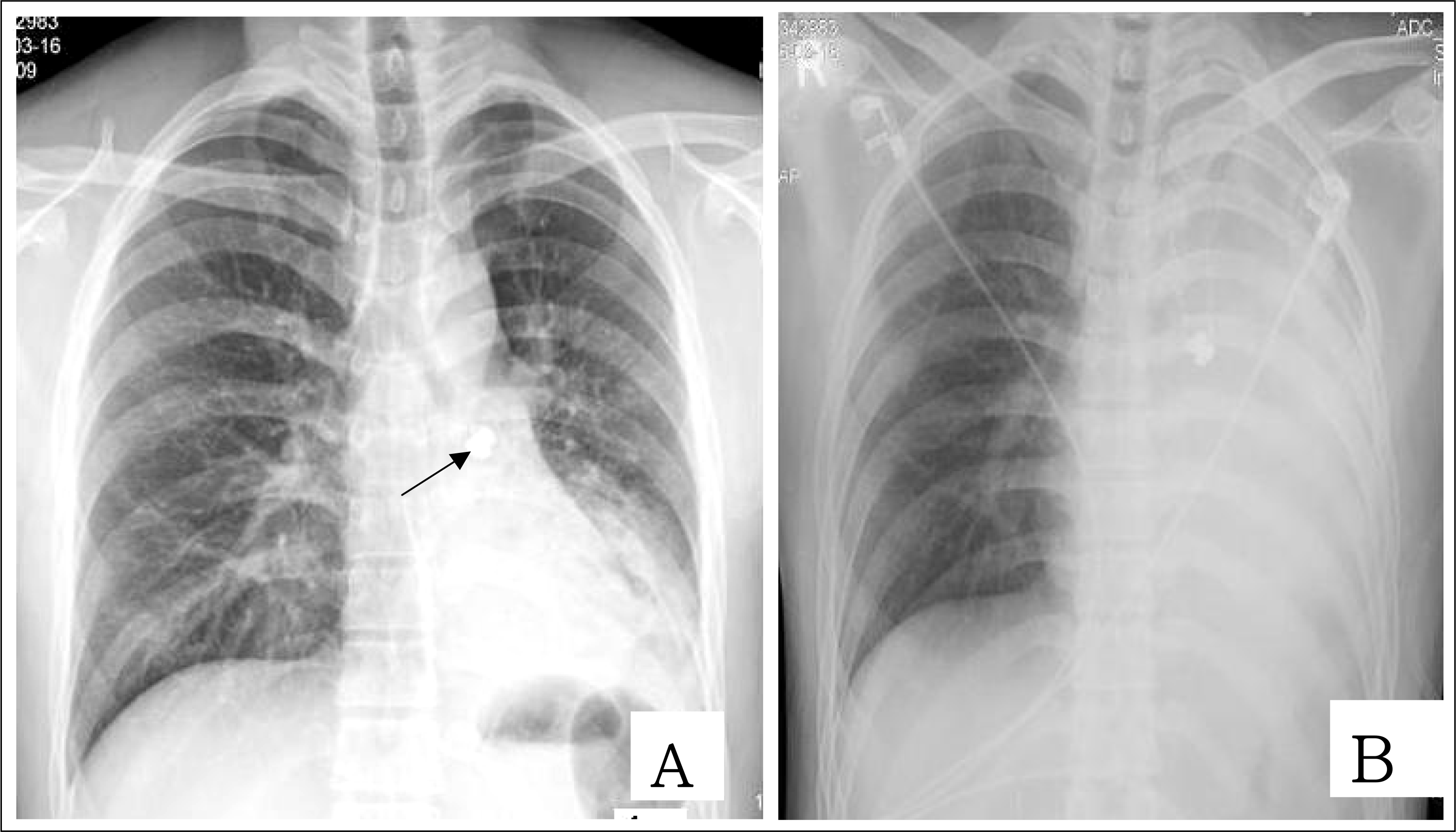 | Figure 1.Chest radiography in a 26-year-old man with hemoptysis and cough. A. The initial chest radiograph shows radio-opaque foreign body in left main bronchus(arrow) and pneumonia infiltration in left lower lung field. B. two days later, Chest X-ray shows nearly complete atelectasis of left lung except some upper lobe. |
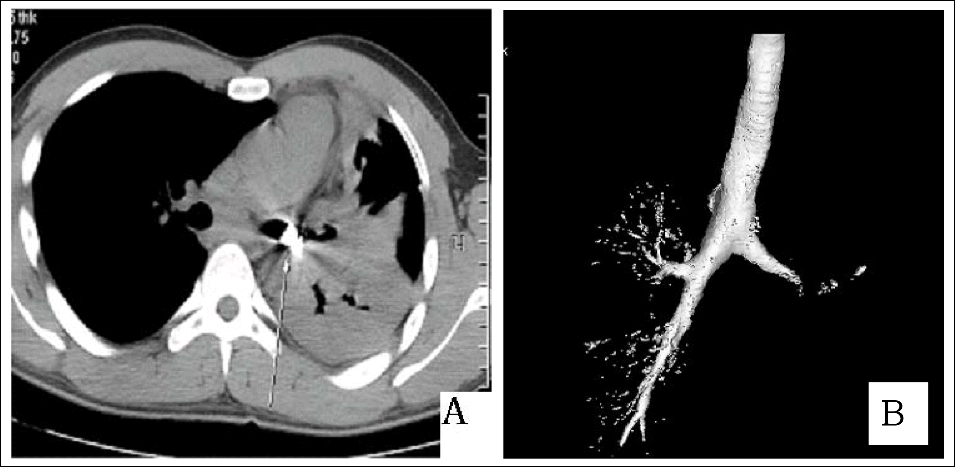 | Figure 2.Chest CT shows metallic foreign body(arrow,A) with nearly complete obstruction in left main bronchus (B). |
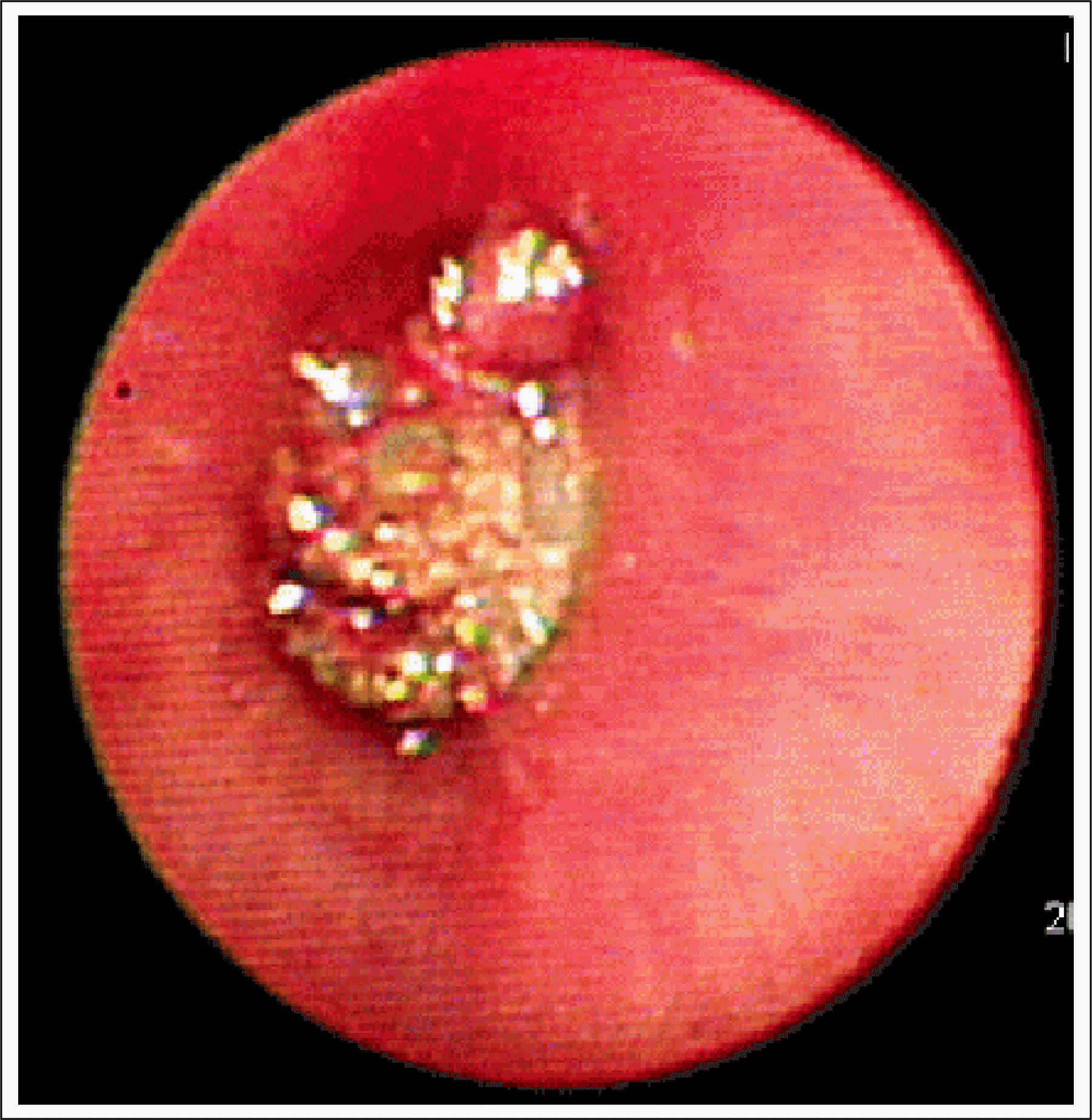 | Figure 3.Bronchoscope shows foreign body covered with mucoid material obstructing left main bronchus. |

 XML Download