

 PDF
PDF ePub
ePub Citation
 Print
Print


Abstract
A multiple primary malignant tumor is a disease mainly encountered in the of the older age groups. Attempts should be made to rule out a second primary malignant neoplasm in the elderly patients with unusual signs and symptoms. We encountered a case of a 67-year-old male with triple primary malignant tumors of the stomach, larynx, and lung. The patient had been treated with a subtotal gastrectomy for early gastric cancer in 1991 and a Laser laryngectomy for the laryngeal squamous cell carcinoma in 2003. In 2005, lung cancer was found with the biopsy revealing an adenosquamous carcinoma. Systemic chemotherapy was performed.
Figures and Tables
Figure 1
(A) Chest PA view shows about 5.2 cm sized thin walled radiolucency (arrow) with ill demarcated osteolytic lesion (arrowhead) in left 6th posterior arc of rib. (B) Lung window of CT scan obtained at the level of azygos arch shows 4.7 cm sized thin walled cavitary mass (arrow) abutting posterior costal pleura in left lower lobe with adjacent focal ground glass attenuated lesion. (C) Mediastinal window of CT scan obtained at the same level of Fig. 2 show thin walled cavity with enhancing posterior costal pleural thickening. Irregular osteolytic lesion (arrow) in posterior arc of left 6th rib is observed. Less than 1 cm sized lymph node enlargement (arrowhead) is seen in left paratracheal area.
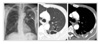
Figure 2
Epiglottis. A(H & E, ×40) & 2B(H&E, ×200). Section disclosed portion shows moderately differentiated squamous cell carcimona.
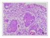
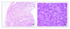
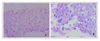
Figure 4
Lung(Adenosquamous carcinoma). A (H & E, ×200) Squamous cell carcinoma shows the proliferation of the squamous cell with atypical chage. B (PAS, ×400). Adenocarcinoma shows cytoplasmic mucin of glandular cell. PAS positive eosinophilic granular material(arrowhead) filled in the grandular cell is shown.
References
1. Kim CK, Chang JW. Multiple primary malignant tumors. J Korean Surg Soc. 1970. 12:63–71.
2. Yoon HK, Kim JP. Multiple primary malignant neoplasm. J Korean Surg Soc. 1984. 26:1–9.
3. Harima M, Yasumoto R, Kawashima H, Asakawa M, Kishimoto T, Maekawa M. A case of asynchronous triple primary malignant tumors of bladder, stomach and lung. Nippon Hinyokika Gakkai Zasshi. 1990. 81:630–633.
4. Hamada Y, Takise A, Uno D, Itoh H, Ichikawa H, Morishta Y. Synchronous primary triple cancers including the lung, stomach, and thyroid. Kyobu Geda. 2000. 53:101–105.
5. Billroth T. Die allgemeine chirugische pathologie and therapies in 51, Vogesusger: in Handbuch fur strudirende and Arzte, 14 Aufl. 1889. Berlin Germany: G Reimer;908.
6. Warren S, Gates P. Multiple primary malignant tumors: a survey of literature and statistical study. Am J Cancer. 1932. 16:1358–1414.
7. Moertel CG, Dockerty MB, Baggenstoss AH. Multiple primary malignant neoplasms: II. tumors of different tissues or organs. Cancer. 1961. 14:231–237.
8. Merscheimer WL, Ringel A, Eisenberg H. Some characteristics of multiple primary cancers. Ann N Y Acad Sci. 1964. 114:896–921.
9. Kwon HM, Chung JB, Kim JH, Chun SI, Cho JK, Park YJ, et al. Multiple primary malignant tumor. Korean J Med. 1987. 33:61–67.
10. Stalker LK, Phillips RB, Pemberton J. Multiple malignant lesions. Surg Gynecol Obstet. 1939. 68:595–602.
11. Slaughter DP. The multiplicity of origin of malignant tumor: collective review. Int Abstr Surg. 1944. 79:89–93.
12. Shin DH, Lee SD, Seo JK. Multiple primary malignant tumors. J Korean Cancer Assoc. 1993. 25:578–585.
13. Burke M. Multiple primary cancers. Am J Cancer. 1936. 27:316–319.
14. Lynch HT, Harris RE, Lynch PM, Guirgis HA, Lynch JF, Bardawil WA. Role of heredity in multiple primary cancer. Cancer. 1977. 40:1849–1854.
15. Yellin A, Hill LR, Benfield JR. Bronchogenic carcinoma associated with upper aerodigestive cancers. J Thorac Cardiovasc Surg. 1986. 91:674–683.
 XML Download