

 PDF
PDF ePub
ePub Citation
 Print
Print


Abstract
Background
PM is known to induce various pulmonary diseases, including asthma, cancer, fibrosis and chronic bronchitis. Despite the epidemiological evidence the pathogenesis of PM-related pulmonary diseases is unclear.
Methods
This study examined the effects of PM exposure on the secretion of TNF-α and IL-1β in the cultured alveolar macrophages. The cultured primary alveolar macrophages were treated with the medium, PM (5~20µg/cm2), LPS (5ng/ml), and PM with LPS for 24h and 48h respectively. ELISA was used to assay the secreted TNF-α and IL-β in the culture medium. Western blotting was used to identify and determine the level of proteins isolated from the culture cells. The cells cultured in the Lab-Tek® chamber slides were stained with immunocytochemical stains.
Results
PM induced TNF-α and IL-1β secretion in the culturing alveolar macrophages, collected from the SPF and inflammatory rats. However, the effects were only dose-dependent in the inflammatory macrophages. When the cells were co-treated with PM and LPS, there was a significant synergistic effect compared with the LPS in the both cell types.
Figures and Tables
Figure 1
Treatment-response relation for TNF-α secretion in the cultured inflammatory BAL cells. Cells were cultured with medium only, LPS (10ng/ml) only and various concentrations of PM (5~20µg/cm2) for 24 hours and 48 hours.
**P<0.01 versus control. Mean ± SEM of four independent experiments per category.
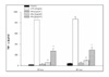
Figure 2
Treatment-response relation for IL-1β secretion in the cultured inflammatory BAL cells. Cells were cultured with medium only, LPS (10ng/ml) only and various concentrations of PM (5~20µg/cm2) for 24 hours and 48 hours.
**P<0.01 versus control. Mean ± SEM of four independent experiments per category.
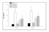
Figure 3
Treatment-response relation for TNF-α secretion in the cultured SPF rat alveolar macrophages at 24 hours and 48 hours. Cells were cultured with medium only (sham control), LPS (5ng/ml), various concentrations of PM (5, 10, 20µg/cm2) and various concentrations of PM with LPS.
*P<0.05, **P<0.01 versus LPS; †P<0.05 versus control. Mean ± SEM of four independent experiments per category (each experiment's n=3)
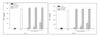
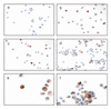
Figure 4
Treatment-response relation for IL-1β secretion in the cultured SPF rat's alveolar macrophages at 24 hours and 48 hours. Cells were cultured with medium only (sham control), LPS (5ng/ml), various concentrations of PM (5, 10, 20µg/cm2) and various concentrations of PM with LPS.
*P<0.05, **P<0.01 versus LPS; †P<0.05 versus control. Mean ± SEM of four independent experiments per category (each experiment's n=3)
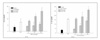
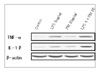
Figure 5
Immunocytochemical stains for TNF-α in the cultured rat alveolar macrophages (× 400). Cells were cultured with (a) medium only, (b) LPS (5ng/ml) only, (c) PM (20µg/cm2) only, and (d) LPS with PM at 6h. (e) LPS only (× 1,000) and (f) LPS with PM (× 1,000) at same incubation time.
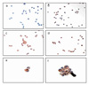
Figure 6
Immunocytochemical stains for IL-1β in the cultured rat alveolar macrophages (× 400). Cells were cultured with (a) medium only, (b) LPS (5ng/ml) only, (c) PM (20µg/cm2) only, and (d) LPS with PM at 6h. (e) LPS only (× 1,000) and (f) LPS with PM(× 1,000) at same incubation time.

References
1. Harre ES, Price PD, Ayrey RB, Toop LJ, Martin IR, Town GI. Respiratory effects of air populution in chronic obstructive pulmonary disease: a three-month prospective study. Thorax. 1997. 52:1040–1044.
2. Lebowitz MD. Epidemiological studies of the respiratory effects of air pollution. Eur Respir J. 1996. 9:1029–1054.
3. Pope CA 3rd, Dockery DW, Spengler JD, Raizenne ME. Respiratory health and PM10 pollution: a daily time series analysis. Am Rev Respir Dis. 1991. 144:668–674.
4. Committee of the Environmental and Occupational Health Assembly of the American Thoracic Society. Health effects of outdoor air pollution: part 2. Am J Respir Crit Care Med. 1996. 153:477–498.
5. Dockery DW, Pope CA 3rd. Acute respiratory effects of particulate air pollution. Annu Rev Public Health. 1994. 15:107–132.
6. Pope CA 3rd, Kanner RE. Acute effects of PM10 pollution on pulmonary function of smokers with mild to moderate COPD. Am Rev Respir Dis. 1993. 147:1336–1340.
7. Emanuel MB. Hay fever, a post industrial revolution epidemic: a history of its growth during the 19th century. Clin Allergy. 1988. 18:295–304.
8. Abbey DE, Hwang BL, Burchette RJ, Vancuren T, Mills PK. Estimated long-term ambient concentrations of PM10 and development of respiratory symptoms in a nonsmoking population. Arch Environ Health. 1995. 50:139–152.
9. Dusseldorp A, Kruize H, Brunekreef B, Hofschreuder P, de Meer G, van Oudvorst AB. Associations of PM10 and airbone iron with respiratory health of adults living near a steel factory. Am J Respir Crit Care Med. 1995. 152:1932–1939.
10. Fels AO, Cohn ZA. The alveolar macrophage. J Appl Physiol. 1986. 60:353–369.
11. Tracey KJ, Cerami A. Metabolic responses to cachectin/TNF. Ann N Y Acad Sci. 1990. 587:325–331.
12. Beutler B, Grau GE. Tumor necrosis factor in the pathogenesis of infectious diseases. Crit Care Med. 1993. 21:S423–S435.
13. Briscoe DM, Cotran RS, Pober JS. Effect of tumor necrosis factor, lipopolysaccharide, and IL-4 on the expression of vascular cell adhesion molecule-1 in vivo: correlation with CD3+T cell infiltrataion. J Immunol. 1992. 149:2954–2960.
14. Gamber JR, Harlan JM, Klebanoff SJ, Vadas MA. Stimulation of the adherence of neutrophils to umbilical vein endothelium by human recombinant tumor necrosis factor. Proc Natl Acad Sci U S A. 1985. 82:8667–8671.
15. Boumpas DT, Chrousos GP, Wilder RL, Cupps TR, Balow JE. Glucocorticoid therapy of immune-mediated diseases: basic and clinical correlates. Ann Intern Med. 1993. 119:1198–1208.
16. Tracy KJ, Fong Y, Hesse DG, Manogue KR, lee AT, Kuo GC, et al. Anti-cachectin/TNF monoclonal antibodies prevent septic shock dmuring lethal bacteraemia. Nature. 1987. 330:662–664.
17. Manel DN, Moore RN, Mergenhagen SE. Macrophages as a sourse of tumoricidal activity (tumor necrotizing factor). Infect Immun. 1980. 30:523–530.
18. Kim KA, Lee DW, Lim Y, Yun IG. Effect of asbestos on fibroblast proliferation of rat. Korean J Occup Environ Med. 1996. 8:392–402.
19. Lim Y, Kim KA, Kim HN, Lee DW, Cho WS, Yun IG. Cytotoxicity and apoptosis by silica, asbestos and man-made mineral fibers. Korean J Occup Environ Med. 1997. 9:641–649.
20. Lim Y, Kim KA, Yun IG. The measurement of IL-1, 8, TNF for the diagnosis of pneumoconiosis. Korean J Occup Environ Med. 1997. 9:17–25.
21. Lim Y, Kim SH, Cho YJ, Kim KA, Oh MW, Lee KH. Silica-inducced oxygen radical ge1neration in alveolar macrophage. Ind Health. 1997. 35:380–387.
22. Vanhee D, Gosset P, Marquette CH, Wallaert B, Lafitte JJ, Gosselin B, et al. Secretion and mRNA expression of TNF alpha and IL-6 in the lungs of pneumocaniosis patients. Am J Respir Crit Med. 1995. 152:298–306.
 XML Download