

 PDF
PDF ePub
ePub Citation
 Print
Print


Abstract
Background :
The incidence of pulmonary tuberculosis has been reducing, but endobronchial tuberculosis continues to be a signigicant heath problem. We performed prospectively bronchoscopy in patients diagnosed with pulmonary tuberculosis in order to evaluate the frequency of endobronchial tuberculosis and its related findings. Follow-up bronchoscopy was also performed after treatment to evaluate the incidence of endobronchial complications such as stenosis and remaining lesions.
Methods :
From January, 1999 to December, 2003, bronchoscopy was performed on patients newly diagnosed with pulmonary tuberculosis.
Results :
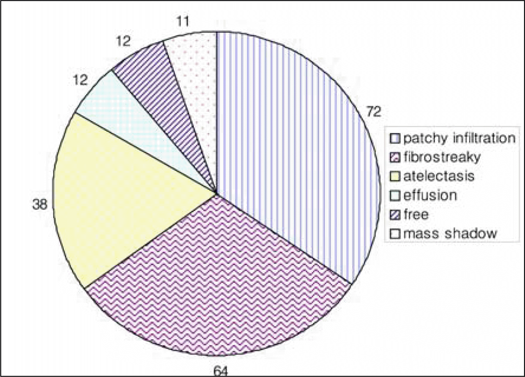
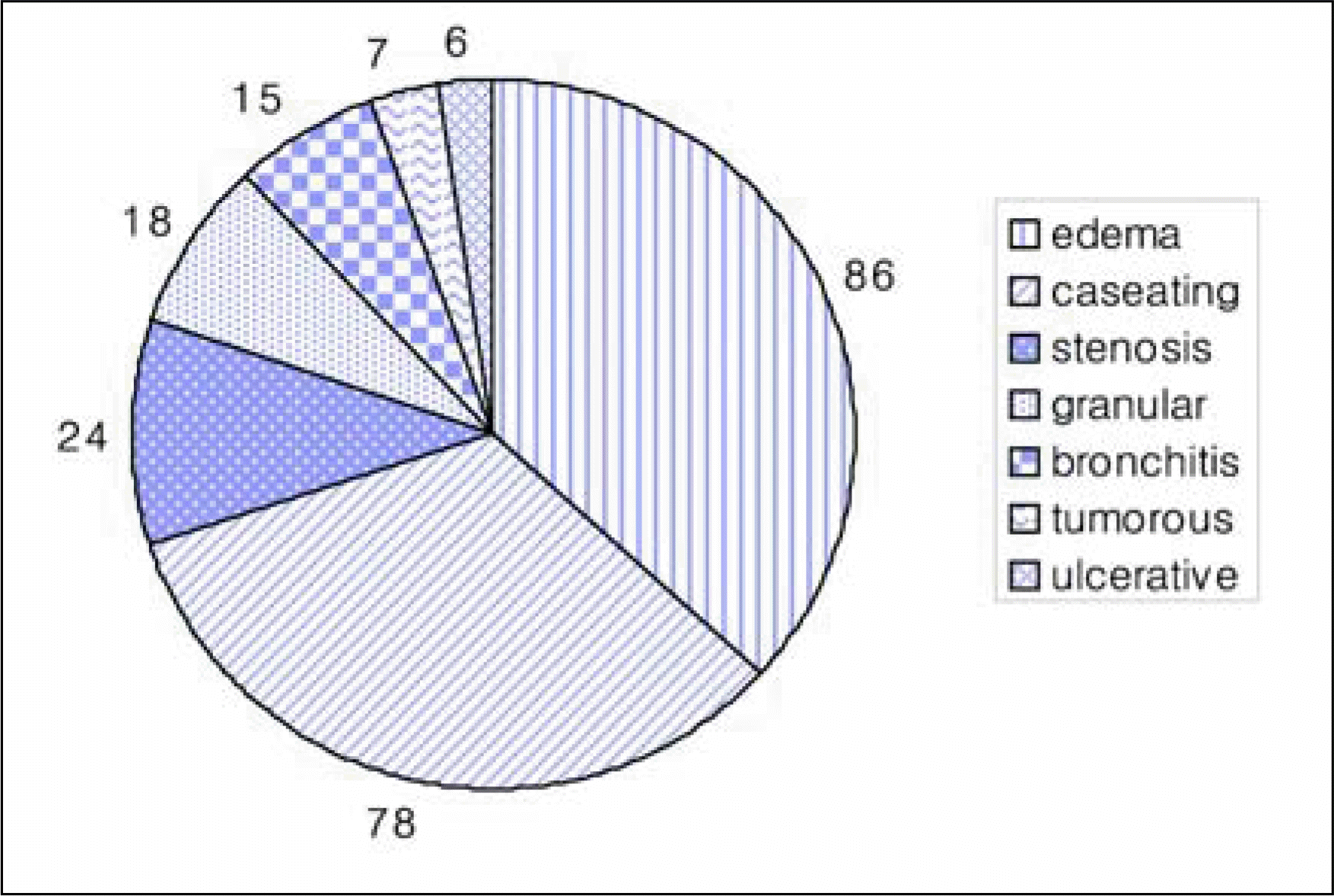
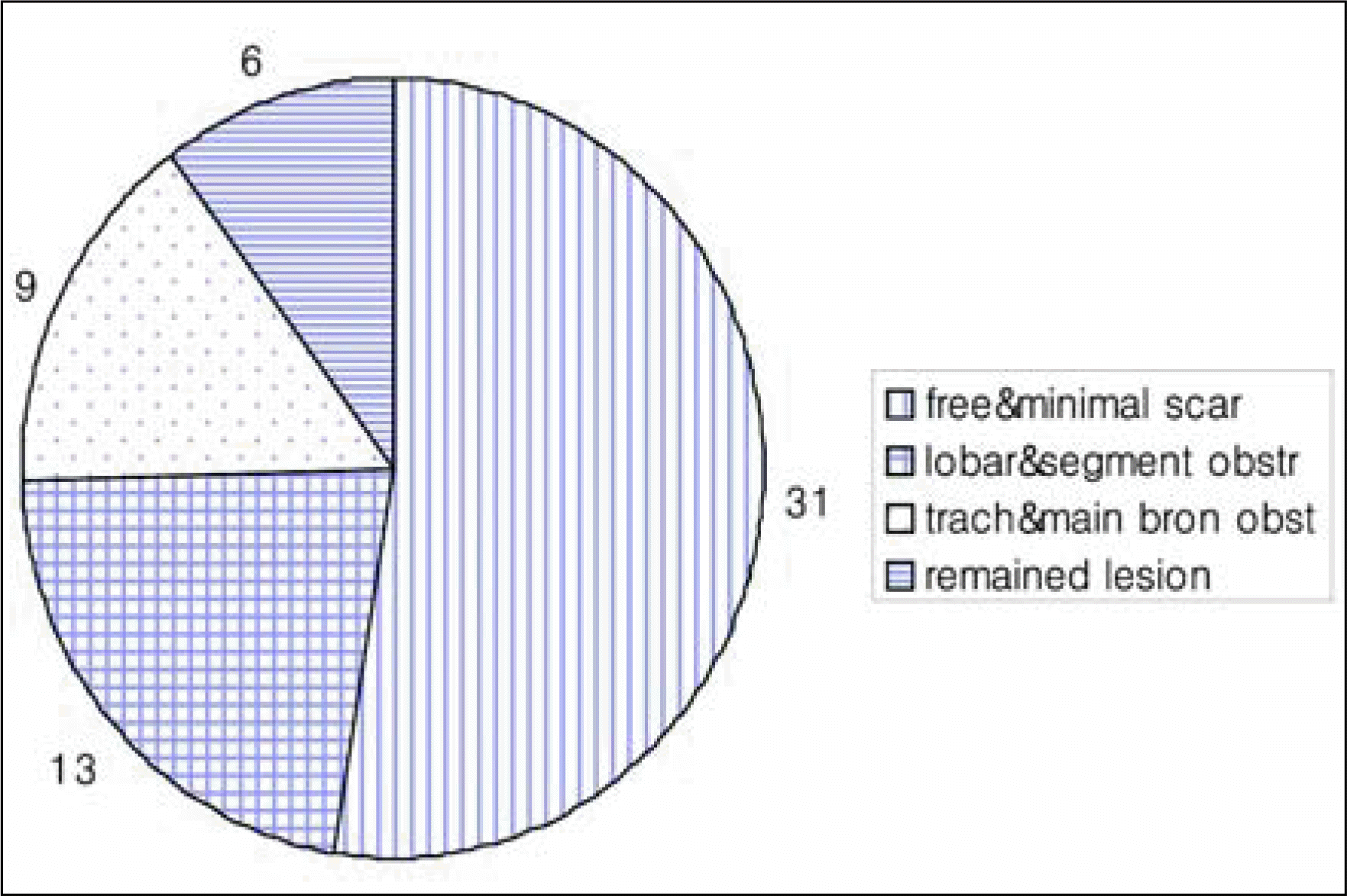
458 patients were enrolled in this study, out of 699 patients with pulmonary tuberculosis from 1999 to 2003. 234(51%) had endobronchial tuberculosis. The frequency was 40.3% in males and 66.3% in females, The most common symptom was nonspecific cough and sputum, and the main radiologiy finding was patchy infiltration. The most common subtype of endobronchial tuberculosis was the edema-hyperemic form. The right lung was involved more frequently than the left, and the left upper lobe was the most commonly involved site. 58 patients underwent follow-up bronchoscopy and most of been cured without major sequels. However, 8 patients had a stenosis of trachea and main bronchus, and 6 patients had still had endobronchial lesions. Therefore the treatment was prolonged for 3 months.
Go to : 
REFERENCES
1.Williams DJ., York EL., Nobert EJ., Sproule BJ. Endobronchial tuberculosis presenting as asthma. Chest. 1988. 93:836–8.

2.Matthews JI., Matarese SL., Carpenter JL. Endobronchial tuberculosis simulating lung cancer. Chest. 1984. 86:642–4.
3.Chung HS., Han SK., Shim YS., Kim KY., Han YC., Kim WS, et al. Balloon dilatation of bronchial stenosis in endobronchial tuberculosis. Tuberc Respir Dis. 1991. 38:236–44.
4.Chung HS., Lee JH., Han SK., Shim YS., Kim KY., Han YC, et al. Classification of endobronchial tuberculosis by the bronchoscopic features. Tuberc Respir Dis. 1991. 38:108–15.
5.WHO. Global tuberculosis control: surveillance, planning, financing: WHO report. 2004.
6.Lew WJ. Tuberculosis surveillance system in Korea. Tuberc Respir Dis. 2000. 48:298–307.
7.Lee JH., Park SS., Lee DH., Shin DH Yang SC., Yoo BM. Endobronchial tuberculosis: clinical and bronchoscopic features in 121 cases. Chest. 1992. 102:990–4.
8.Kim SY., Suhr JW., Shin KS., Jeong SS., Park SG., Kim AK, et al. Endobronchial tuberculosis in patients with pulmonary tuberculosis. Tuberc Respir Dis. 1996. 43:138–46.
9.Park EJ., Kim MO., Yang SC., Sohn JW., Yoon HJ., Shin DH, et al. Clinical and bronchoscopic features of 280 patients with endobronchial tuberculosis: 1990-2001. Korean J Med. 2003. 64:284–92.
10.Ahn CM., Kim HJ., Hwang ES., Kim SK., Lee WY. A clinical study of 61 cases of tuberculous tracheobronchitis. Tuberc Respir Dis. 1991. 38:340–6.
11.Hirata S. Tracheobrochial tuberculosis observed from the chest X-ray findings and its pathogenesis. Kek-kaku. 1989. 64:319–27.
12.Jokinen K., Palva T., Nuutinen J. Bronchial findings in pulmonary tuberculosis. Clin Otolaryngol Allied Sci. 1977. 2:139–48.
13.Song JH., Han SK., Heo IM. Clinical study of endobronchial tuberculosis. Tuberc Respir Dis. 1985. 32:276–82.
14.Chung HS., Lee JH. Bronchoscopic assessment of the evolution of Endobronchial tuberculosis. Chest. 2000. 117:385–92.
15.Toppet M., Malfroot A., Derde MP., Toppet V., Spehl M., Dad I. Corticosteroid in primary tuberculosis with bronchial obstruction. Arch Dis Chil. 1990. 65:1222–6.
16.Chan HS., Pang JA. Effect of corticosteroid on deterioration of endobronchial tuberculosis during chemotherapy. Chest. 1989. 96:1195–6.
Go to :
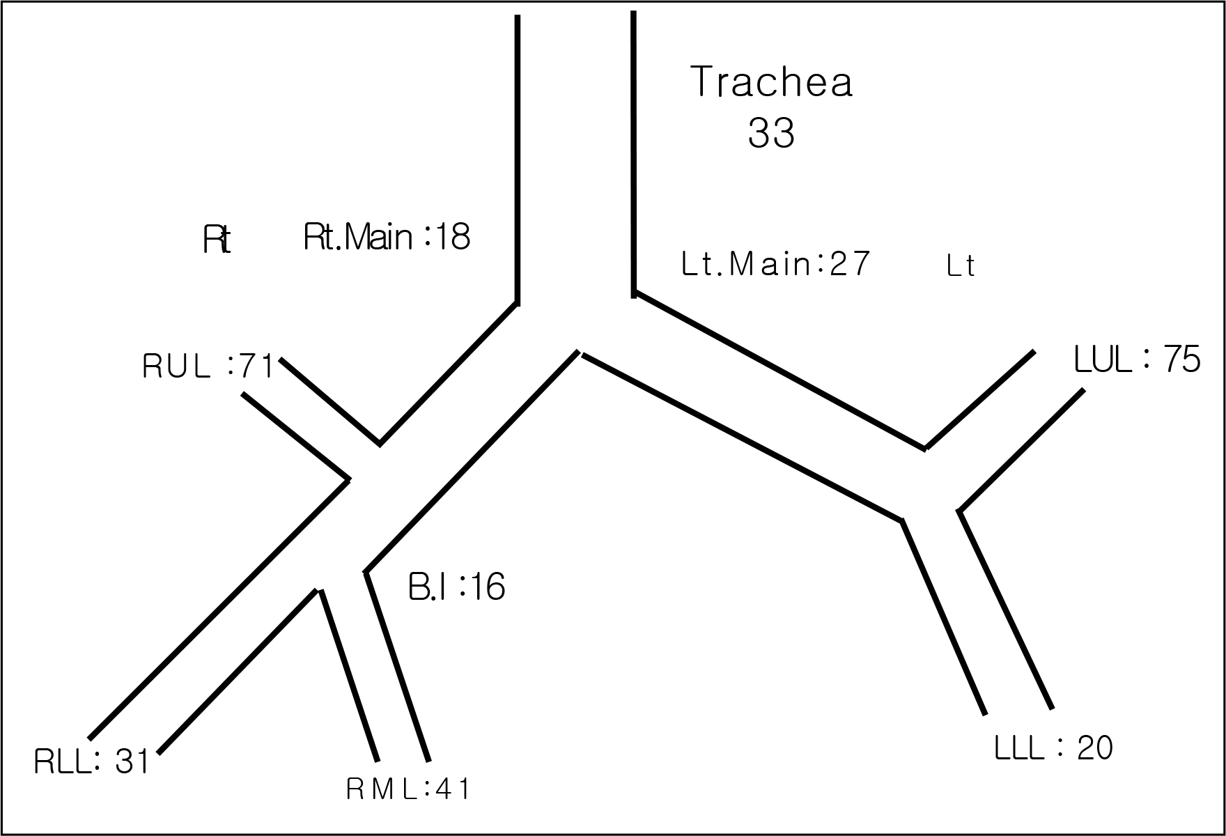
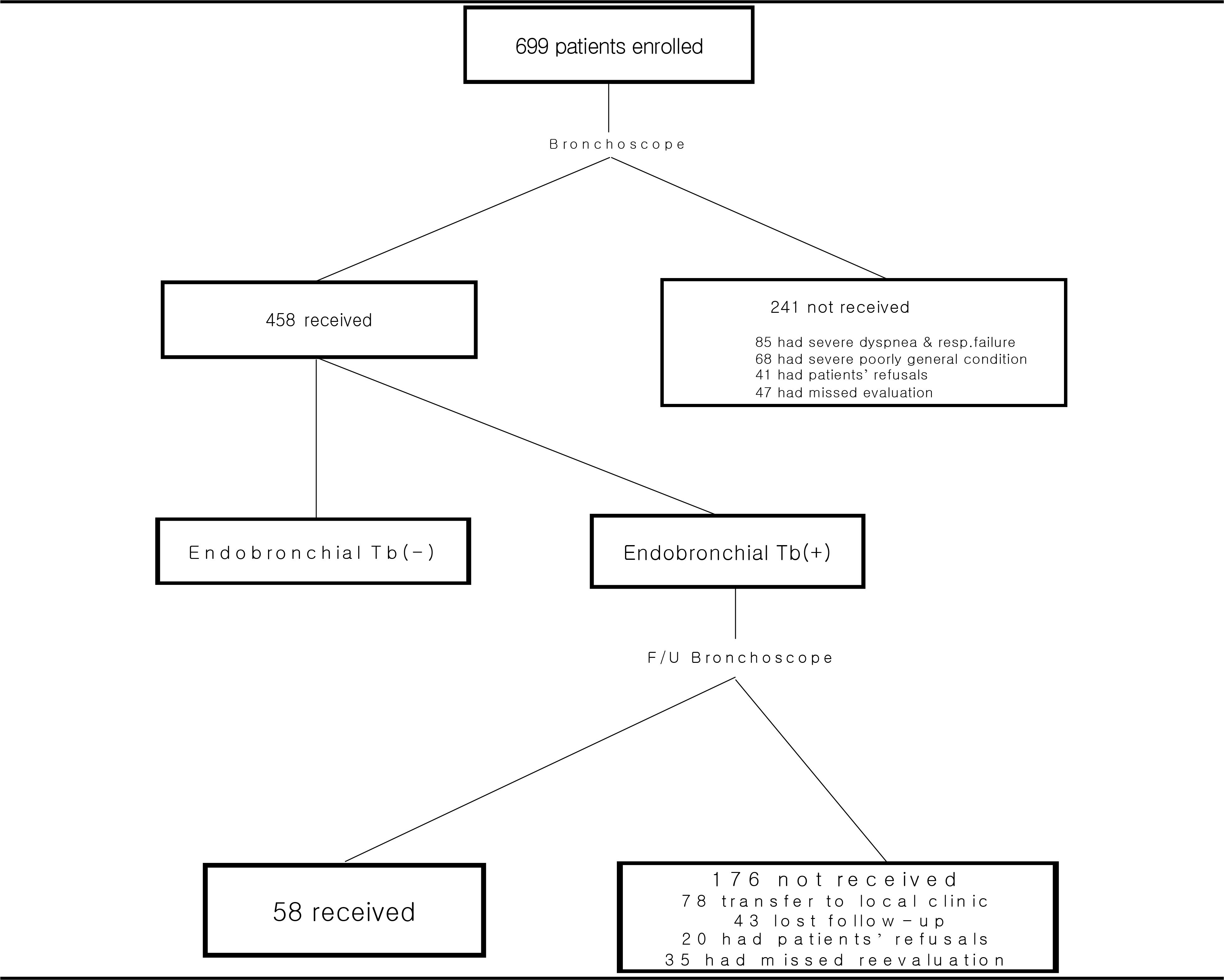
Table 2
: Age distribution of the cases
| N. Of Endobronch ial TB | Age | ||||||
|---|---|---|---|---|---|---|---|
| <20 | 20- | 30- | 40- | 50- | 60- | >70 | |
| No | 13 | 38 | 34 | 33 | 39 | 37 | 24 |
| Yes | 14 | 46 | 23 | 30 | 27 | 38 | 43 |
| % | 51.8 | 54.7 | 40.3 | 47.6 | 40.9 | 50.6 | 64.1 |
 XML Download