

 PDF
PDF ePub
ePub Citation
 Print
Print


Abstract
Background
Particulate matters (PM) when inhaled is known to induce pulmonary diseases including asthma and chronic bronchitis when inhaled. Despite the epidemiological proofevidence, the pathogenesis of PM-related pulmonary diseases is unclearremain poorly understood.
Methods
Primary alveolar macrophages were harvested from the SPF and inflammatory rats by bronchioalveolar lavage (BAL). The cultured primary alveolar macrophages were treated with the medium only, PM only (5~40µg/cm2), LPS (5ng/ml) only, and PM with LPS for 24 and 48 hours. The level of secreted nitric oxide (NO) was assayed from the cultured medium by using the Griess reaction. The cultured cells were utilized for the western blotting against the inducible nitric oxide synthase (iNOS) proteins. Immunocyto- chemical staining against the iNOS and NT-proteins were performed in cells that cultured in the Lab-Tek® chamber slide after treatments.
Results
The PM that utilizein this experiments induced NO formation with iNOS expression in the cultured SPF and inflammatory rats alveolar macrophages, by itself. When the cells were co-treated with PM and LPS, there was a statistically significant synergistic effect on NO formation and iNOS expression over the LPS effect. The cells from the sham control showed minimal immunoreactivity for the NT-proteins. Significantly higher quantities of NT-proteins were detected in the PM and PM with LPS co-treated cells than from the sham control.
Figures and Tables
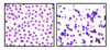 | Figure 1Composition and morphology of BAL cells (×400). Diff-Quick stained alveolar macrophages from the SPF rat (a) and from the inflammatory rat (b).
|
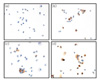
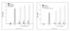 | Figure 2
Figure 2A. Treatment-response relation for NO2- formation in cultured SPF rat alveolar macrophages at 24 hours. Cells were cultured with medium only (sham control), LPS (5ng/ml), various concentrations of PM (5, 10, 20µg/cm2) and various concentrations of PM with LPS.
*P<0.05 versus LPS; †P<0.01 versus control. Mean ± SEM of four independent experiments per category (each experiment's n=3).
Figure 2B. Treatment-response relation for NO2- formation in cultured SPF rat alveolar macrophages at 48 hours. Cells were cultured with medium only (sham control), LPS (5ng/ml), various concentrations of PM (5, 10, 20µg/cm2) and various concentrations of PM with LPS.
**P<0.01 versus LPS; †P<0.01 versus control. Mean ± SEM of four independent experiments per category (each experiment's n=3).
|
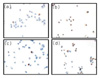
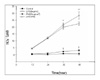 | Figure 3Time-response relation for NO2- formation in cultured SPF rat alveolar macrophages. Cells were cultured with medium only (control), LPS only, PM only and PM with LPS for 12-48 hours.
*P<0.05, **P<0.01 versus LPS; †P<0.01 versus control. Mean ± SEM of four independent experiments per category.
|
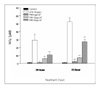 | Figure 4Treatment-response relation for NO2- formation in cultured inflammatory BAL cells. Cells were cultured with medium only, LPS (10ng/ml) only and various concentrations of PM (5~20µg/cm2) for 24 hours and 48 hours.
*P<0.05, **P<0.01 versus control. Mean ± SEM of four independent experiments per category.
|
 | Figure 5Immunocytochemical stain for iNOS in the cultured rat alveolar macrophages (×400). Cells were cultured with (a) medium only, (b) LPS (5ng/ml) only, (c) PM(20µg/cm2) only and (d) LPS with PM for 48 hours.
|
 | Figure 6Immunocytochemical stain for nitrotyrosiliated-protein (NP) in cultured rat alveolar macrophages (×400). Cells were cultured with (a) medium, (b) LPS (5ng/ml), (c) PM (20µg/cm2) and (d) LPS with PM (20µg/cm2) for 48 hours.
|
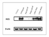 | Figure 7Western blot for iNOS in cultured rat alveolar macrophages. Cells were cultured with medium only, LPS only, PM only and PM with LPS for 24 hours. The level of β-actin was similar in all of the tested samples.
|

References
1. Salma I, Willy M, Eva ZP, Gyula A. Comprehensive characteri sation of atmospheric aerosols in Budapest, Hungary: physicochemical properties of inorganic species. Atmos Environ. 2001. 35:4367–4378.
2. Schwartz J, Dockery DW, Neas LM. Is daily mortality associated specifically with fine particles? J Air Waste Manag Assoc. 1996. 46:927–939.
3. Hung YC, Guh JH, Cheng ZJ, Chang YL, Hwang TL, Liao CH, et al. Inhibition of the expression of inducible nitric oxide synthase and cyclooxygenase-2 in macrophages by 7HQ derivatives: involvement of lkappaB-alpha stabilization. Eur J Pharmacol. 2001. 418:133–139.
4. Liang YC, Huang YT, Tsai SH, Lin-Shiau SY, Chen CF, Lin JK. Suppression of inducible cycloxygenase and inducible nitric oxide synthase by apigenin and related flavonoids in mouse macrophages. Carcinogenesis. 1999. 20:1945–1952.
5. Cui S, Reichner JS, Mateo RB, Albina JE. Activated murine macrophages induce apoptosis in tumor cells through nitric oxide-dependent or independent mechanisms. Cancer Res. 1994. 54:2462–2467.
6. Zhang X, Alley EW, Russell SW, Morrison DC. Necessity and sufficiency of beta interferon for nitric oxide production in mouse peritoneal macrophages. Infect Immun. 1994. 62:33–40.
7. Vazquez-Torres A, Jones-Carson J, Balish E. Nitric oxide production does not directly increase macrophage candidacidal activity. Infect Immun. 1995. 63:1142–1144.
8. Morin AM, Stanboli A. Nitric oxide synthase localization in cultured cerebrovascular endothelium during mitosis. Exp Cell Res. 1994. 211:183–188.
9. Adams LB, Franzblau SG, Vavrin Z, Hibbsand JB Jr, Krahenbuhl JL. L-arginine-dependent macrophage effector functions inhibit metabolic activity of Mycobacterium leprae. J Immunol. 1991. 147:1642–1646.
10. Nathan CF. Secretory products of macrophages. J Clin Invest. 1987. 79:319–326.
11. Salvemini D, Radziszewski W, Korbut R, Vane J. The use of oxyhaemoglobin to explore the events underlying inhibition of platelet aggregation induced by NO or NO-donors. Br J Pharmacol. 1990. 101:991–995.
12. Zhang J, Megaridis CM. Soot suppression by ferrocene in laminar ethylene/air nonpremixed flames. Combust Flame. 1996. 105:528–540.
13. Harrison RM, Yin J. Particulate matter in the atmosphere: which particle properties are important forits effects on health? Sci Total Environ. 2000. 249:85–101.
14. Ito T, Ikeda M, Yamasaki H, Sagai M, Tomita T. Peroxynitrite formation by diesel exhaust particles in alveolar cells: links to pulmonary inflammation. Environ Toxicol Pharmacol. 2000. 9:1–8.
15. Pandya RJ, Solomon G, Kinner A, Balmes JR. Diesel exhaust and asthma: hypotheses and molecular mechanisms of action. Environ Health Persect. 2002. 110:Suppl 1. 103–112.
16. Dong CC, Yin XJ, Ma JX, Millecchia L, Wu ZX, Barger MW, et al. Effects of diesel exhaust particles on allergic reactions and airway responsiveness in ovalbumin-sensitized brown Norway rats. Toxicol Sci. 2005. 88:202–212.
17. Choe N, Tanaka S, Kagan E. Asbestos fibers and interleukin-1 upregulate the formation of reactive nitrogen species in rat pleural mesothelial cells. Am J Respir Cell Mol Biol. 1998. 19:226–236.
 XML Download