

 PDF
PDF ePub
ePub Citation
 Print
Print


Abstract
Background
Pulmonary tuberculosis is frequently accompanied with complications such as bronchiectasis, cavities, fibrosis and a deterioration of the lung function. However, there is little information available on the pathogenesis of these complications in pulmonary tuberculosis. Among the many factors involving in tissue remodeling, transforming growth factor-β1 (TGF-β1) is a potent stimulus of the extracellular matrix fomation and a mediator of potential relevance for airway wall remodeling. Therefore, this study examined the relationship between the radiological changes and the TGF-β1 level in patients with pulmonary tuberculosis.
Methods
Serum and bronchoalveolar lavage fluid (BALF) were collected from total of 35 patients before treating them for active pulmonary tuberculosis, and the TGF-β1 levels were measured using an enzyme-linked immunosorbent assay (ELISA). The BALF levels were recalculated as the epithelial lining fluid (ELF) levels using the albumin method. pulmonary function test (PFT) and high resolution computed tomography (HRCT) were performed before and after treatment.
Results
There was a strong correlation between the serum TGF-β1 level and the presence of cavities (r=0.404, p=0.006), even though the BAL TGF-β1 level showed a weak correlation with complications. In addition, there was no correlation between the TGF-β1 levels before treatment and the changes in the PFT and HRCT during treatment.
Figures and Tables
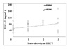
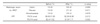
Table 2
Differences of TGF-β1 levels in BALF and serum before treatment, subdivided into two groups according to the changes HRCT score and PFT after treatment
Data are presensented as median, range. BALF levels were recalculated as epithelial lining fluid levels by the albumin method. Abbreviation: BALF, bronchoalveolar lavage fluid; HRCT, high resolution computed tomography; PFT, pulmonary function test; FVC, forced vital capacity; FEV1, forced expiratory volume in one second
![]()
References
1. Murray CJ, Styblo K, Rouillon A. Tuberculosis in developing countries: burden, intervention and cost. Bull Int Union Tuberc Lung Dis. 1990. 65:6–24.
2. Blobe GC, Schiemann WP, Lodish HF. Role of transforming growth factor beta in human disease. N Engl J Med. 2000. 342:1350–1358.
3. Massague J. The transforming growth factor-β family. Annu Rev Cell Biol. 1990. 6:597–641.
4. Grainger DJ, Kemp PR, Liu AC, Lawn RM, Metcalfe JC. Activation of transforming growth factor-beta is inhibited in transgenic apolipoprotein(a) mice. Nature. 1994. 370:460–462.
5. Border WA, Noble NA. Transforming growth factor beta in tissue fibrosis. N Engl J Med. 1994. 331:1286–1292.
6. Roche WR, Beasley R, Williams JH, Holgate ST. Subepithelial fibrosis in the bronchi of asthmatics. Lancet. 1989. 1:520–524.
7. Ameglio F, Casarini M, Capoluongo E, Mattia P, Puglisi G, Giosue S. Post-treatment changes of six cytokines in active pulmonary tuberculosis: differences between patients with stable or increased fibrosis. Int J Tuberc Lung Dis. 2005. 9:98–104.
8. Casarini M, Ameglio F, Alemanno L, Zangrilli P, Mattia P, Paone G, et al. Cytokine levels correlate with a radiologic score in active pulmonary tuberculosis. Am J Respir Crit Care Med. 1999. 159:143–148.
9. Kim KU, Lee SJ, Lee JH, Cho WH, Jung KS, Joe JH, et al. The correlation between bronchostenosis and changes in the levels of interferon-γ and transforming growth factor-β during the treatment in patients with endobronchial tuberculosis. Tuberc Respir Dis. 2005. 58:18–24.
10. Ryu YJ, Kim YJ, Kwon JM, Na YJ, Jung YJ, Seoh JY, et al. Circulating cytokine levels and changes during the treatment in patients with active tuberculosis in Korea. Tuberc Respir Dis. 2003. 55:140–153.
11. Jang AS, Park SW, Ahn MH, Park JS, Kim DJ, Lee JH, et al. Impact of circulating TGF-β and IL-10 on T cell cytokines in patients with asthma and tuberculosis. J Korean Med Sci. 2006. 21:30–34.
12. Rennard SI, Basset G, Lecossier D, O'Donnell KM, Pinkston P, Martin PG, et al. Estimation of volume of epithelial lining fluid recovered by lavage using urea as marker of dilution. J Appl Physiol. 1986. 60:532–538.
13. Raviglione MC, Snider DE Jr, Kochi A. Global epidemiology of tuberculosis: morbidity and mortality of a worldwide epidemic. JAMA. 1995. 273:220–226.
14. Fletcher HA, Owiafe P, Jeffries D, Hill P, Rook GA, Zumla A, et al. Increased expression of mRNA encoding interleukin (IL)-4 and its splice variant IL-4delta2 in cells from contacts of Mycobacterium tuberculosis, in the absence of in vitro stimulation. Immunology. 2004. 112:669–673.
15. de la Barrera S, Aleman M, Musella R, Schierloh P, Pasquinelli V, Garcia V, et al. IL-10 down-regulates costimulatory molecules on Mycobacterium tuberculosis-pulsed macrophages and impairs the lytic activity of CD4 and CD8 CTL in tuberculosis patients. Clin Exp Immunol. 2004. 138:128–138.
16. Bai X, Wilson SE, Chmura K, Feldman NE, Chan ED. Morphometric analysis of Th(1) and Th(2) cytokine expression in human pulmonary tuberculosis. Tuberculosis. 2004. 84:375–385.
17. Hirsch CS, Yoneda T, Averill L, Ellner JJ, Toossi Z. Enhancement of intracellular growth of Mycobacterium tuberculosis in human monocytes by transforming growth factor-β. J Infect Dis. 1994. 170:1229–1237.
18. Toossi Z, Gogate P, Shiratsuchi H, Young T, Ellner JJ. Enhanced production of TGFβ by blood monocytes from patients with active tuberculosis and presence of TGFβ in tuberculous granulomatous lung lesions. J Immunol. 1995. 154:465–473.
19. Rich EA. Pulmonary immune response to Mycobacterium tuberculosis and human immunodeficiency virus. Infect Agent Dis. 1996. 5:108–118.
20. Im JG, Itoh H, Shim YS, Lee JH, Ahn J, Han MC, et al. Pulmonary tuberculosis: CT findings-early active disease and sequential change with antituberculous therapy. Radiology. 1993. 186:653–660.
21. Murata K, Itoh H, Todo G, Kanaoka M, Noma S, Itoh T, et al. Centrilobular lesions of the lung: demonstration by high-resolution CT and pathologic correlation. Radiology. 1986. 161:641–645.
22. Tozkoparan E, Deniz O, Ciftci F, Bozkanat E, Bicak M, Mutlu H, et al. The roles of HRCT and clinical parameters in assessing activity of suspected smear negative pulmonary tuberculosis. Arch Med Res. 2005. 36:166–170.
23. Altin R, Savranlar A, Kart L, Mahmutyazicioglu K, Ozdemir H, Akdag B, et al. Presence and HRCT quantification of bronchiectasis in coal workers. Eur J Radiol. 2004. 52:157–163.
24. Wang YH, Lin AS, Lai YF, Chao TY, Liu JW, Ko SF. The high value of high-resolution computed tomography in predicting the activity of pulmonary tuberculosis. Int J Tuberc Lung Dis. 2003. 7:563–568.
25. Yabuuchi H, Murayama S, Murakami J, Sakai S, Hashiguchi N, Soeda H, et al. Correlation of immunologic status with high-resolution CT and distributions of pulmonary tuberculosis. Acta Radiol. 2002. 43:44–47.
 XML Download