

 PDF
PDF ePub
ePub Citation
 Print
Print


INTRODUCTION
Regular resistance exercise is an excellent strategy that enhances physical fitness, including muscle strength and skeletal muscle hypertrophy [123]. Although the optimal resistance exercise protocol for promoting muscular strength is unknown, it is common for resistance-trained men to perform resistance exercise to skeletal muscle hypertrophy. Resistance exercise provides mechanical tension, which induces skeletal muscle overload [45]. It has been postulated that resistance exercise–induced chronic skeletal muscle damage in men contributes to the triggering of skeletal muscle dysfunction via increase in markers of skeletal muscle damage [6]. These skeletal muscle damage markers, including creatine kinase (CK), creatine kinase–myocardial band (CK-MB), troponin(s), B-type natriuretic peptide (BNP), lactate dehydrogenase (LDH) [78910], and myoglobin, play a role in skeletal muscle soreness through tears in supportive connective tissue [11], sarcolemma [12], basal lamina [13], z-disk [14], and in structures that injure contractile elements and the cytoskeleton [15]. Furthermore, interventions that decrease markers support attenuation of skeletal muscle damage, increase in physical function, and maintenance of the intracellular anabolic metabolism for training adaptation process. Toward this end, different approaches have been tried to recognize the best novel supplementation strategy that can protect against increased skeletal muscle damage markers in resistance-trained men.
Various nutritional approaches [16], in particular plant-origin products [17], enhance skeletal muscle function [18] and anabolic response to resistance exercise [19]. Although dietary supplementation has been shown to increase muscle strength, it has not been determined whether dietary supplementation with resistance training (RT) may protect against increase in skeletal muscle damage markers in resistance-trained men. Furthermore, the importance of this study is highlighted by the recent rise in strategies for prevention of skeletal muscle damage using supplementation in resistance-trained men [20]. In particular, ursolic acid (UA), a pentacyclic triterpenoid carboxylic acid, is found in various plants, edible vegetables, and medicinal herbs. Its biological activities are widely recognized, including its anti-oxidant, anti-inflammatory, and anti-hyperlipidemic effects [2122]. Recently, UA has been reported to increase skeletal muscle mass [23], reverse ischemia-induced cardiac dysfunction in mouse cardiac myocytes [24], and improve cardiac failure in animals [25]. However, the effects of UA on skeletal muscle damage markers in resistance-trained men are yet to be investigated. Thus, we aimed to determine the role of UA supplementation on the levels of markers of skeletal muscle damage in resistance-trained men.
METHODS
Participants
Sixteen healthy resistance-trained male volunteers (mean age, 33.00±1.30 years; mean body weight, 85.14±3.16 kg) were enrolled in the study. All participants were experienced resistance-trained (RT) athletes who had consistently trained under personalized skeletal muscle hypertrophy programs for 3 years. To further verify the effect of UA on skeletal muscle damage in resistance-trained men, participants without injury who performed RT at least three times per week to sustain skeletal muscle hypertrophy with repetition range, as previously described in the literature, were recruited [26]. Participants with chronic diseases such as cardiovascular diseases, hypertension, diabetes, or obesity diagnosed 6 months before the study were excluded. Participants were randomly divided into two groups: RT (control group) and RT+UA (intervention group). The intervention was performed for 8 weeks. All participants was given informed consent before participation in the study, and ethical approval was given by the Institutional Review Board at Pusan National University.
RT protocol
The RT program was designed by a professional strength and conditioning specialist. The specialist determined the 1 repetition maximum (1 RM) intensity for each subject before the study, as previously described [27]. All subjects trained 6 times per week and adopted the program to become familiar with all exercise sessions. The program was started in the early evening after the participants returned from work. Participants performed the RT program for 8 weeks, consisting 26 exercise types (13 upper-body and 13 lower-body training exercises). Every exercise included 60% to 80% of 1 RM, and all five sets were completed with 60 to 90 s inter-set rest, as previously described [27].
UA supplementation
The participants took one 150 mg UA capsule (Labrada, Houston, TX, USA) after each meal, for a total of 3 capsules/day (450 mg in total), for 8 weeks. The supplementation protocols were given in detail in our previous study [27]. We monitored the dietary pattern of the subjects via cellular phone or via laboratory visits during the study.
Body composition and blood parameters
Body composition and blood parameters were measured before and after the 8-week intervention period. Body composition was measured using a multi-frequency electrical impedance analyzer (X-scan Plus II, Jawon Medical, Seoul, Korea). Blood samples were obtained from the antecubital vein after 10 hours of fasting. The blood samples were centrifuged at 1,500 g and 4℃ for 15 min and frozen at -80℃ until analysis. CK, LDH, and CK-MB levels were measured using an automated analyzer from Hoffman-LaRoche (Basel, Switzerland). Cortisol level was measured using an automated analyzer from Hitachi (Tokyo, Japan). myoglobin level was measured using an automated analyzer from Beckman Coulter (Brea, CA, USA). BNP level was measured using enzyme-linked immunosorbent assay kits from Biosite Inc. (San Diego, CA, USA) [28].
Statistics analysis
All data were expressed as mean±standard error (SEM) using SPSS version 22.0 (IBM, Armonk, NY, USA). To determine the mean difference between groups, the data were analyzed using two-way analysis of variance with repeated measurements (group [RT and RT+UA] by time [before and after 8 weeks]). If the interaction (time x group) was found to be significant, within-group comparisons were made using paired t-test. Statistical significance was set at p<0.05.
RESULTS
Characteristics of participants
Participants were assigned to either the RT group (n=8) or the RT+UA group (n=8) for the 8-week intervention period. The baseline characteristics of the study participants are presented in Table 1. There were no significant differences in the baseline characteristics of the participants between both groups. Body weight and body fat percentage slightly decreased in both groups after 8 weeks, but were not statistically significant. On the contrary, lean body mass was slightly increased in both groups after 8 weeks, but without statistical significance.
Makers of skeletal muscle damage
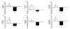
The changes in the levels of skeletal muscle damage markers from baseline to after 8 weeks are presented in Table 2. In the RT group, there were no significant changes in any parameters before and after 8 weeks, whereas for the RT+UA group, there was a significant treatment-by-time interaction for BNP, CK, CK-MB, cortisol, LDH, and myoblobin (p<0.05). In addition, in the RT+UA group, UA supplementation significantly decreased the levels of BNP, CK, CK-MB, and LDH (p<0.05). The changes in all parameters were significantly (p<0.05) different between groups (Fig. 1).
DISCUSSION
In the present study, we found that RT with UA supplementation in resistance-trained men caused a decline in the levels of skeletal muscle damage markers, such as BNP, CK, CK-MB, and LDH. However, we did not find significant reduction in body weight or body fat percentage or increase in skeletal muscle mass. These findings suggest that UA supplementation has beneficial effects on attenuating the increase in skeletal muscle damage markers in resistance-trained men during the 8-week RT.
Regular RT enhances skeletal muscle mass and muscular strength, which sustains physical fitness [29]. RT leads to successful skeletal muscle hypertrophy, but it also causes increase in skeletal muscle damage markers and reduction in skeletal muscle regenerative factors [3031]. Thus, it is important to maintain the balance of these parameters in resistance-trained men. Additionally, the use of these programs without concomitant nutritional support can lead to skeletal damage and soreness, which can have an effect on increased CK in blood and block the recovery of skeletal muscle damage and function [32]. Based on these previous results, we hypothesized that the participants who underwent regular, high-intensity RT would have a higher level of skeletal muscle damage markers during the 8 weeks. Unexpectedly, we found that the level of skeletal muscle damage markers, such as CK (39.73%), BNP (19.08%), CK-MB (2.58%), cortisol (14.66%), LDH (8.23%), and myoglobin (10.16%), showed a slight tendency to increase with RT, but statistical significance was not reached. It is possible that in some of the previous studies resistance-trained men, who always performed high intensity RT, have sustained increase in skeletal damage markers, compared with untrained men [33]. However, other study reported that an increase in CK levels in blood damages the skeletal muscle cell structure [34] and leads to a decrease in exercise performance after high RT periods [35]. These findings suggest that the RT protocol cannot possibly affect higher levels of these markers in resistance-trained men compared with untrained men. In addition, the discrepancy between the present and previous studies might be due to difference in RT intensity, and duration of training in resistance-trained men. Thus, further studies are necessary to elucidate the influence of RT protocols on the exercise-induced release of skeletal muscle damage markers to understand its role in the physiological response to RT protocols in resistance-trained men.
Recently, growing evidences suggest that UA is beneficial for the improvement of energy expenditure and skeletal muscle function through the activation of protein synthesis and inhibition of skeletal muscle atrophy [363738]. Additionally, the rationale behind our selection of UA was the recent strong finding based on the level of biomarkers of cardiac and liver damage in disease rodent models [3940]. However, whether UA supplementation affects release of skeletal muscle damage markers during RT in resistance-trained men is unclear. As expected, we found that RT with UA supplementation in resistance-trained men promoted a decline in the level of skeletal muscle damage markers, such as serum BNP, CK, CK-MB, and LDH. These findings suggest that the decrease in the levels of these markers induced by UA may lead to the recovery of the skeletal muscle damage markers during RT in resistance-trained men. It is important to note that UA supplementation is considered to contribute to decrease in skeletal muscle damage markers. A similar study by Radhiga et al. [40] suggested that UA could decrease CK, CK-MB, and LDH in rats with cardiac infarct. In addition, Bakhtiari et al. [36] reported that UA promoted skeletal muscle regeneration by differentiation of satellite cells. These results suggest that UA exhibited protective effect against increased skeletal damage markers and degradation of regeneration factors including satellite cells in resistance-trained men. This study, therefore, is the first to report that UA supplementation effectively suppresses skeletal muscle damage markers in resistance-trained men. Our results provide a compelling evidence that UA can be a potential dietary method for inhibition of skeletal muscle damage markers during RT in resistance-trained men, with in-depth molecular mechanisms requiring further investigation.
The presented results have some limitations. First, the sample size observed in this study was relatively small. Second, we only studied healthy resistance-trained men. Future study on UA supplementation with and without RT should be performed in participants with chronic diseases such as skeletal muscle atrophy, aging, obesity, and diabetes. Third, follow-up studies are necessary to determine the relationship between skeletal muscle damage markers and skeletal muscle function. Finally, the duration of high-intensity exercise was short.
In conclusion, the present study revealed that UA supplementation inhibited the skeletal muscle damage markers during RT in resistance-trained men, hence suggesting that it will be an alternative therapy against skeletal muscle damage after RT.
 XML Download