Abstract
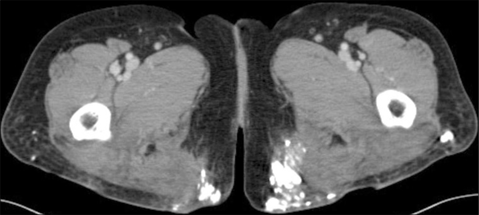
The calcinosis, dystrophic soft tissue calcification, occurs in damaged or devitalized tissues normal calcium/phosphorus metabolism. It is the subcutaneous tissues of connective tissues disease - primarily systemic lupus erythematosus, scleroderma, or dermatomyositis - and may involve a relatively localized area. The calcinotic accumulations may result in muscle atrophy, joint contractures, and skin ulceration complicated by recurrent episodes of local inflammation and infection. Calcinosis may be the source of both pain and disability in connective tissue disease patients. While various therapeutic modality have been used, no treatment has convincingly prevented or reduced calcinosis. We report two cases of calcinosis cutis combined with rheumatic disease.
REFERENCES
1). 박진현: 심영학: 배선우: 이광훈: 조휘율: 김호근. 전신성홍반성낭창에동반된범발성연부조직석회증. 대한내과학회지. 1989. 37:427–32.
2). Boulman N., Slobodin G., Rozenbaum M., Rosner I. Calcinosis in rheumatic diseases. Semin Arthritis Rheum. 2005. 34:805–12.

3). Halverson PB., Ryan LM. Arthritis associated with calcium-containing crystals. Klippel JH, editor. ed.Primer on the rheumatic diseases. 12th ed.p. 299306. Atlanta, GA: Arthritis Foundation;2001.
4). Tan AWH., Ng HJ., Ang P., Goh YT. Extensive calcinosis cutis in relapsed acute lymphoblastic leukaemia. Ann Acad Med Singapore. 2004. 33:107–9.
5). Qadri SRM., Choksey MS., Shad A. Tumoural calcinosis of the cervical spine: case report, pathogenesis and differential diagnosis. Br J Neurosurg. 2005. 19:185–90.

6). Guermazi A., Grigoryan M., Cordoliani F., Kerob D. Unusually diffuse idiopathic calcinosis cutis. Clin Rheumatol. 2007. 26:268–70.

7). Terranova M., Amato L., Palleschi GM., Massi D., Fabbri P. A case of idiopathic calcinosis universalis. Acta Derm Venereol. 2005. 85:189–90.

8). Wilmer A., Magro M. Calciphylaxis: emerging concepts in prevention, diagnosis, and treatment. Seminars in Dialysis. 2002. 15:172–86.

9). Angelis M., Wong LL., Myers SA., Wong LM. Calciphylaxis in patients on hemodialysis: a prevalence study. Surgery. 1997. 122:1083–90.

10). Marzano AV., Kolesnikova LV., Gasparini G., Alessi E. Dystrophic calcinosis cutis in subacute lupus. Dermatology. 1999. 198:90–2.

11). Whyte MP. Extraskeleta (ectopic) calcification and ossification. In:. Favus MJ, Christakos S, Kleerekoper M, Shane E, Gagel R, Langman CB, Stewart AF, editors. eds.Primer on the metabolic bone diseases and disorders of mineral metabolism. 2nd ed.p. 386–95. New York, Raven Press;1993.
12). Cukierman T., Elinav E., Korem M., Chajek-Shaul T. Low dose warfarin treatment for calcinosis in patients with systemic sclerosis. Ann Rheum Dis. 2004. 63:1341–3.

13). Abdallah-Lotf M., Grasland A., Vinceneux P., Sigal-Grinberg M. Regression of cutis calcinosis with diltiazem in adult dermatomyositis. Eur J Dermatol. 2005. 15:102–4.
14). Ambler GR., Chaitow J., Rogers M., McDonald DW., Ouvrier RA. Rapid improvement of calcinosis in juvenile dermatomyositis with alendronate therapy J Rheumatol. 2005. 32:1837–9.
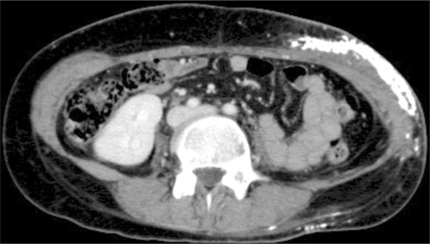
Fig. 1.
Abdomen-pelvis CT at diagnosis, Case 1. There are extensive calcifications and fascial thickening involving left side abdominal wall, subcutaneous layer predominantly.


 PDF
PDF ePub
ePub Citation
 Print
Print


 XML Download