

 PDF
PDF ePub
ePub Citation
 Print
Print


Abstract
Developmental coxa vara is a rare disease and the symptoms do not appear at birth, but rather, they appear at the age of walking. Clinically, the symptoms include a waddling gait, limb length discrepancy and frequent weariness. Developmental coxa vara is sometimes associated with skeletal dysplasia. Especially, it is associated with spondylometaphyseal dysplsia and the vertebral bodies and long bones are affected. The authors report here on diagnosing and treating this rare disease and we review the relevant literatures.
Figures and Tables
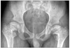
Figure 1
(A) A radiograph shows coxa vara with decreased neck-shaft angle and increased Hilgenreiner-epiphyseal angle (H-E angle). (B) A radio graph shows spondylometaphyseal dysplasia characterized with platyspondyly. (C) There is a marked sclerosis, flaring of the metaphyseal end of distal tibia and fibula. (D) Irregular areas of metaphyses also present corner fracture on medial aspect of proximal tibia.
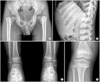
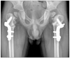
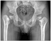
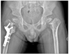
Figure 3
Anteroposterior radiograph taken 2 years after subtrochanteric valgus osteotomy shows the recurrence of developmental coxa vara on the right hip.
References
1. Morrissy RT, Weinstein SL. Lovell and Winter's pediatric orthopaedics. 2000. 5th ed. Philadelphia: Lippincott-Raven;1035–1044.
2. Sutcliffe J. Metaphyseal dysostosis. Ann of Radiol. 1966. 9:215–223.
3. Kozlowski K, Bellemore MC. Spondylo-metaphyseal dysplasia of Sutcliffe type. Br J Radiol. 1989. 62:862–864.

4. Song KS, Joh BJ. Coxa vara with spondylometaphyseal dysplasia - case report -. J Korean Orthop Assoc. 1996. 31:988–991.
5. Herkowitz HN, Grafin SR, Balderston RA, et al. Rothman-Simeone The spine. 1992. 4th ed. Philadelphia: Saunders;303–314.
6. Davies RW, Hall CM, Apley AG. Atlas of skeletal dysplasias. 1985. Edinburgh London Melbourne and New York: Churchill Livingstone;171–177.
7. Lee SH, Suh SW, Rha KW, Jung HI, Jo JH. A case of spondylometaphyseal dysplasia (Kozlowski type). J Korean Orthop Assoc. 1997. 32:768–772.
8. Langer LO Jr, Brill PW, Ozonoff MB, et al. Spondylometaphyseal dysplasia, corner fracture type: a heritable condition associated with coxa vara. Radiology. 1990. 175:761–766.
9. Carroll K, Coleman S, Stevens PM. Coxa vara: surgical outcomes of valgus osteotomies. J Pediatr Orthop. 1997. 17:220–224.
10. Krakow D, Vriens J, Camacho N, et al. Mutations in the gene encoding the calcium-permeable ion channel TRPV4 produce spondylometaphyseal dysplasia, Kozlowski type and metatropic dysplasia. Am J Hum Genet. 2009. 84:307–315.
 XML Download