

 PDF
PDF Citation
 Print
Print


INTRODUCTION
Human papillomavirus (HPV) is the causative agent for cervical cancer [1]. This heterogenous virus family includes more than 200 genotypes [2]; among which, more than 40 HPV types can easily spread through genital tract [3]. Fourteen HPV genotypes (HPV 16, 18, 31, 33, 35, 39, 45, 51, 52, 56, 58, 59, 66, and 68) are considered pathogenic or “high-risk” for causing the development of cervical cancer [45]. Although most sexually active females become infected with HPV once in their lifetime [6], less than 10% of women become persistently infected [7], and it is the ‘persistent’ infection with a high-risk genotype HPV that contributes to cervical cancer development [789].
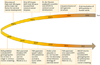
In recent decades, great advances have been made in understanding the molecular biology of HPV, and the importance of HPV genotyping, a method that identifies specific HPV genotypes, has become more widely recognized (Fig. 1). HPV particles were first visualized in the mid-1900s, and in the late 1990s, high-risk HPV genotypes were revealed to be the main risk factor for development of cervical cancer. During the last two decades, a number of HPV genotyping tests have been developed, and three types of vaccines designed to prevent HPV infection have been approved by the U.S. Food and Drug Administration (FDA). HPV genotyping is essential for preventing cervical cancer, and investigations concerning the therapeutic use of genotyping tests are in progress. To elucidate the role of HPV genotyping in cervical cancer, the following subjects are addressed in this review: (1) clinically important issues in HPV virology; (2) the current application of HPV genotyping in clinical medicine; and (3) potential future uses for HPV genotyping.
CLINICALLY IMPORTANT HPV VIROLOGY
Knowledge of HPV virology is essential for understanding cervical cancer development. The structure of the HPV genome was characterized in in 1965 (Fig. 1) [10], and this information greatly contributed to understanding the role of HPV in cervical cancer [111213].
1. HPV genome
The HPV is a small (50 to 55 nm in diameter and ~8 kb in length) double stranded DNA virus that exhibits tropism toward epithelial cells, and infects the skin and mucosa (Fig. 2) [1415]. It consists of icosahedral capsid composed of 72 capsomers that have shapes resembling five-pointed stars. A virus genome exists inside the capsid, and harbors eight partially overlapping open reading frames. The genome is divided into three regions: an early region (E), late region (L), and a long control region (LCR) [516].
Fig. 2
The human papillomavirus (HPV) genome and a schematic view of HPV-mediated cervical cancer progression. HPV consists of six early genes (E), two late genes, and a long control region (L). HPV virions infect the cervical basal epithelial cells and contribute to cervical cancer development. CIN, cervical intraepithelial neoplasia; CIS, carcinoma in situ; LCR, long coding region.
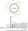
The early region of the genome encodes early genes E1, E2, E4, E5, E6, and E7 (Fig. 2), which are involved in viral replication, transcription regulation, and oncogenesis (Table 1) [5]. The products of early genes E6 and E7 are oncoproteins that play important roles in cancer progression. The oncoprotein from E6 binds to tumor suppressor gene p53, where it impairs DNA repair, inhibits apoptosis, and destabilizes the chromosome. Oncoprotein E7 binds to retinoblastoma (Rb) protein, which is a well known tumor suppressor, and promotes dysregulation of the cell cycle. Additionally, oncoprotein E7 also interacts with several cellular proteins other than Rb protein, and is involved in apoptosis inhibition and evasion of immune surveillance functions [51517].
Table 1
Functions of human papillomavirus genes

The late region of the genome encodes structural proteins L1 and L2 (Fig. 2). These proteins comprise the capsid protein, protect the viral genome located inside (Table 1), and are expressed in the upper layer of the epithelium [5]. Additionally, L1 protein can assemble itself into empty capsid-like structures, and its immunogenicity is similar to that of infectious virions. Hence, current vaccines used to prevent HPV infection include L1 protein as a main constituent [1819]. L2 protein is necessary for allowing viral entry into cells, the transport of viral components into the nucleus, and their binding with DNA. L2 protein evokes production of a broad spectrum of neutralizing antibodies against different types of HPV. These antibodies are more cross-reactive between HPV genotypes when compared to the antibodies provoked by L1. As a result, L2 protein is considered to be an important component of future vaccines [20].
Unlike the other two regions (early and late regions), the long control region (LCR) is a noncoding upstream regulatory region located between E6 and L1 (Fig. 2). It contains a core promoter sequence, as well as enhancer and silencer sequences, and is necessary for viral replication and transcription (Table 1). The size and nucleotide composition of the LCR display considerable variation among different HPV genotypes [521].
2. HPV classification
HPV belongs to the family papillomaviridae, and its classification is clinically significant for the following reasons: (1) only one specific HPV genus is associated with cervical cancer; (2) the pathogenicity of HPV varies according to genotype. HPV is grouped into five genera (alpha, beta, gamma, mu, and nu), and the genus Alphapapillomavirus includes HPV genotypes that infect both genital and oral mucosa [14]. Additionally, HPV can be classified based on its L1 genome sequence, type, intratypic lineage, and sublineage. Different types, intratypic lineages, and sublineages of HPV are defined by having L1 sequences that differ by at least 10%, more than 1%, and less than 1%, respectively [322]. HPV can also be grouped into categories of “high-risk” and “low-risk” based on their oncogenic potential. Among 14 high-risk HPV genotypes (HPV 16, 18, 31, 33, 35, 39, 45, 51, 52, 56, 58, 59, 66, and 68), the two most common (HPV 16 and 18) are the causative factors for 71% of cervical cancers [23]. Two low-risk HPV genotypes (HPV 6 and 11) contribute to the formation of genital warts, most of which require treatment [14].
3. Life cycle of HPV
The transformation of HPV infected cells to cancer cells is a multi-step process [924]. HPV infects basal cells located in the epithelial transformation zone (Fig. 2). This transformation zone exists between the stratified squamous epithelium of the ectocervix and the columnar epithelium of the endocervix, and provides an entry site for HPV [25]. The viral replication process begins shortly after the virus enters a host cell [26]. Initial viral replication is tightly linked to the epithelial cell differentiation cycle. HPV infects only dividing basal epithelial cells; thus HPV DNA replicates only when basal cell DNA is replicated [27]. Genes E1 and E2 are required for the maintenance of viral genomes in host cells, as they serve as the initial sites for replication of viral DNA, and also recruit cellular DNA polymerase needed for replication [28]. E6 and E7 oncoproteins act to enhance cellular proliferation, resulting in increased numbers of infected cells and infectious virions [2829]. In summary, carcinogenesis is a multi-step process, not only because viral genes take various actions to transform a normal cervical cell into a cervical cancer cell, but also because cervical epithelial tissue progresses through phases of being normal epithelium, cervical intraepithelial neoplasia tissue (CIN; CIN 1, CIN 2, and CIN 3), and carcinoma in situ, when developing into cervical cancer (Fig. 2). Overexpression of viral genes results in the transformation of HPV infected cells to malignant cells [172729].
CURRENT APPLICATION OF HPV GENOTYPING IN CLINICAL FIELD
1. Cervical cancer screening
Cervical sampling is used to detect HPV infection after amplifying expression of the viral genome or mRNA [30], and several FDA-approved HPV tests are commercially available (Table 2) [31323334353637]. Traditional Papanicolaou (Pap) screening [38] was first implemented 50 years ago. In 2012, American Cancer Society guidelines for the early detection of cervical cancer began including HPV DNA testing as a method to be used in conjunction with cytology or part of a triage of tests that can be employed to further investigate abnormal cytology findings [39]. This recommended use of HPV DNA testing has been incorporated into current clinical practice. According to the National Comprehensive Cancer Network guideline, co-testing with the Pap and HPV tests is a first-line cervical cancer screening method, and it is recommended that women aged 30 to 65 years have these tests performed every 5 years [40]. Its main advantage is that it provides improved sensitivity for detecting CIN 2 lesions [41]. The best method for managing women with normal cytology findings but who test positive for HPV has been a subject for debate. The current recommendation is to perform either a follow-up test 12 months later or genotyping of HPV 16 and 18. If the genotyping of HPV 16 and 18 is ‘negative,’ co-testing after 12 months is recommended; if it is ‘positive,’ further examination with colposcopy is recommended [42].
Table 2
Food and Drug Administration-approved HPV tests

| Molecular target | Principle | Name | Remark | |
|---|---|---|---|---|
| DNA based assay | ||||
| High-risk HPV DNA test | Full genome | Hybridization | Hybrid capture 2 (HC2) HPV DNA test | Not designed for genotyping individual HPV types |
| L1 | Invader assay | Cervista HPV HR test | High sensitivity in the detection of CIN 2+ [31] | |
| High-risk HPV DNA tests with partial genotyping for the main high-risk HPV types | L1 | Real-time PCR | Cobas 4800 HPV test | Approved for HPV primary screening [32] |
| Hybridization | Cervista HPV 16/18 test | Low false positive rate with high sensitivity and specificity to genotyping HPV 16 and 18 [3334] | ||
| L1 | Real-time PCR assay | Abbot RealTime HR HPV test | High specificity with no cross reaction with low-risk HPV types [35] | |
| mRNA based assay | ||||
| High-risk HPV mRNA test | E6/E7 | Transcription mediated amplification | APTIMA HPV test | No cross reaction with low-risk HPV types [36] |
| High-risk HPV mRNA test with partial genotyping for the main high-risk HPV types | E6/E7 | Transcription mediated amplification | APTIMA HPV 16, 18/45 test | Includes HPV 45 to identify more women at risk for adenocarcinoma, in addition to HPV 16 and 18 [37] |
The National Cancer Institute conducted the ASC-US-LSIL Triage study (ALTS), and the results supported the use of triage for women with atypical squamous cells of undetermined significance (ASC-US) [43]. When used for diagnosing women aged >21 years with ASC-US, the triage method identified 96% of CIN 3+ cases, and only 56% of cases were referred for colposcopy; indicating that triage testing has sensitivity comparable to that of colposcopy. In addition, the ALTS study led to the conclusion that the low-risk HPV types are less likely to contribute cervical cancer development, thus testing for such is not clinically valuable. Subsequently, triage platforms with genotyping of high-risk HPV types and/ or genotyping of HPV 16 and 18 have been established with success [4445]. However, the prevalence of high-risk HPV infections was too high to permit an accurate evaluation of triage for diagnosing low-grade squamous intraepithelial lesions (LSIL).
HPV testing is used in follow-up after CIN treatment in order to monitor possible recurrence [4647] and a recent data reported type-specific HPV genotyping improves the prediction of CIN recurrence as compared to HPV testing [48]. It is known that ~10% of women treated for CIN 3+ develop residual/ recurrent disease [49]. High sensitivity is essential in detecting HPV infection in residual/recurrent disease [50] and a recent meta-analysis reported that HPV testing improved the sensitivity (85% to 97%) as compared to the conventional cytology [51].
FUTURE CLINICAL APPLICATION OF HPV GENOTYPING
1. New era of cervical cancer screening
As guidelines for cervical cancer screening continue to evolve, HPV genotyping has assumed a significant role in newly published recommendations. In 2014, the FDA approved an HPV test for use in primary screening for cervical cancer [52]. Additionally, the Society of Gynecologic Oncology and the American Society for Colposcopy and Cervical Pathology recently released interim guidelines that endorsed screening with an HPV test alone (without cytology) every 3 years for women ≥25 years old [32]. Based on previous data [53], the guidelines recommended using different clinical approaches for managing patients infected with different HPV genotypes identified by HPV genotyping (Supplementary Fig. 1). Patients positive for HPV 16/18 would undergo colposcopy, while patients infected with any of 12 other high-risk HPV types would be tested by reflex cytology. Although issues regarding patient preferences and reimbursement remain unresolved, most health practitioners support the effectiveness of HPV genotyping as a primary screening modality [54]. Recent findings suggest that the use of HPV genotype testing as a primary screening tool will increase, and thus play a more important role in cervical cancer prevention.
An HPV test is more sensitive than cytology for detecting cervical precancerous cervical lesions, and less affected by the individual who collects the specimen [5355565758]. Therefore, HPV genotyping using self-collected samples might be an option to reduce costs and increase patient participation in HPV screening programs. Studies comparing the results of HPV tests performed using self-and clinician-collected samples showed equivalent HPV genotype distributions and prevalence [5960]. HPV genotyping using self-collected samples was feasible and well accepted, and showed sensitivity and specificity comparable to those achieved using clinician-collected samples [61]. Moreover, self-testing detected precancerous cervical lesions even earlier than cytology [62]. Home-based HPV testing is a good alternative not only for people residing in developed countries, but also for people living in developing countries where clinics are not easily accessible. Previous observations showed that a home-based HPV genotyping test identified sufficient numbers of women at risk for cervical cancer to produce a reduction in morality [63]. However, the test displayed limited specificity, and thus might require the use of additional triage tests (e.g., triage with cytology or methylation-marker testing) when used to confirm HPV positive results [64].
2. Therapeutic HPV vaccines
HPV genotyping is required to select individuals eligible to receive vaccines being studied for safety and efficacy in clinical trials [6566]. Vaccines that prevent HPV infection provide little protection in women with a pre-existing HPV infection [6768]; therefore, therapeutic vaccines that might control an existing infection are currently under investigation. Moreover, therapeutic vaccines should not only manage HPV-related lesions, but also establish a systemic immunological memory to help prevent disease recurrence [69]. Many of the therapeutic vaccines currently being studied contain the E6/E7 oncogenes of specific high-risk HPV genotypes (particularly HPV 16 and 18), and work by inducing a robust cellular immune response that eradicates HPV-related lesions [65707172]. For example, Kim et al. [65] developed a therapeutic vaccine using HPV 16-specific CD8+ cytotoxic T-lymphocyte responses that stimulates the expansion of CD8α+ lymphoid dentritic cells and facilitated the expression of HPV antigen through the major histocompatibility complex class I pathway. Therefore, HPV genotyping must be conducted to identify individuals with high-risk HPV types (particularly HPV 16 and 18) who would benefit from receiving a therapeutic vaccine.
CONCLUSIONS
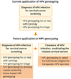
This review examined the role of HPV genotyping in the prevention and treatment of a cervical cancer or precancerous lesion. We elucidated the structure of the HPV genome, the functions of each gene during the HPV life-cycle, and how they relate to development of cervical cancer (Table 1). The history of HPV research shows that the field has achieved great scientific advances during the past two decades (Fig. 1). And although, previous data reported that HPV genotyping has limitations in its suboptimal specificity [73], it has significantly reduced the burden of HPV-related cervical lesions. HPV genotyping is changing from being a supporting method used to help prevent cervical cancer to a main method that also assists in managing pre-existing cervical lesions (Fig. 3). Although persistent infection with a high-risk HPV genotype is known to be a major carcinogenic factor, the various high-risk HPV genotypes have different carcinogenic potentials [23]. Therefore, an understanding of the genotype-specific aspects of HPV infection would facilitate the development of better strategies to prevent and manage cervical cancer.
Fig. 3
Schematic representation showing changes in the application of human papillomavirus (HPV) genotyping. HPV genotyping is currently perceived as a supporting method used in cervical cancer screening, but it will become a main method in the future. The yellow box denotes future applications of HPV genotyping in cervical cancer prevention and management.
 XML Download