Journal List > J Dent Anesth Pain Med > v.18(4) > 1100263

Nath, Daneshmand, Sizemore, Guo, and Enciso: Efficacy of corticosteroids for postoperative endodontic pain: A systematic review and meta-analysis

Abstract

This systematic review aimed to analyze the efficacy of corticosteroid premedication compared to placebo or no treatment to reduce postoperative pain in endodontic patients. Randomized controlled trials (RCTs) assessing corticosteroids via oral, intramuscular, subperiosteal, intraligamentary or intracanal route compared to passive or active placebo, or no treatment were included. Four databases were searched: PubMed, Web of Science, Cochrane Library and Embase up to 2/21/2018. Risk of bias was assessed with Cochrane Risk of bias tool. Fourteen RCTs with 1,462 generally healthy adults in need of endodontic treatment were included. 50% of the studies were at unclear risk and 50% at high risk of bias. Meta-analysis showed Visual Analog Scale (VAS) pain at 4–6 hours after Inferior Alveolar Nerve Block (IANB) was significantly lower by 21 points (0–100 scale) in the corticosteroid group compared to the control group (95% CI −35 to −7; P = 0.003), however this difference was not statistically significant after 24 hours (P = 0.116). The route of administration was oral and intraligament injection. Patients who received corticosteroids prior to IANB were 70.7% more likely to have none or mild pain 4–8 hours after treatment (P = 0.001) and 13.5% more likely 24 hours after IANB (P = 0.013) than patients in the control group. In conclusion, corticosteroid administration (oral or intraligamental) may clinically reduce the level of postoperative pain at 4–8 hours after IANB, however the quality of the evidence was low/moderate due to risk of bias and heterogeneity. Further studies are recommended.

INTRODUCTION

The International Association for the Study of Pain [1] in 1994, has defined pain as an “unpleasant sensory and emotional experience associated with actual or potential tissue damage, or described in terms of such damage” [1]. The complex mechanisms of pain are still being actively researched and elucidated [2]. The incidence of significant postoperative pain in endodontic patients is reported to be between 3–58% [3]. The wide variation can be linked to differences in study design, technique protocol, pain measurement, operator experience, and pre-existing mitigating factors such as inflammation and infection [2]. Pain origin associated with endodontic therapy primarily includes bacterial (either from an abscess, or introduced during treatment, swelling, instrumentation), chemical (irrigants/intracanal medication) or hyper-occlusion[4].
For reduction of pain during endodontic therapy clinicians employ diverse non-pharmacologic measures such as occlusal reduction, incision and drainage, trephination, careful extirpation, cleaning and obturation, as well as non-traditional measures such as hypnosis [5]. However, to control and minimize the post-operative pain, analgesics (over the counter, or prescribed), opioids or corticosteroids may be required [6]. The use of a corticosteroid to reduce pre-operative, intra-operative and post-operative endodontic pain was described as early as 1956 by Stewart [7]. There are no conclusive guidelines on the ideal route of administration or the corticosteroid of choice. A prior literature review recommends that the steroid be delivered at least a few hours prior to treatment [8].
Pharmacodynamics of drugs are complex, having its effect at the receptor level in cell membranes, in the mitochondria, and nuclei [9]. Though the underlying mechanism of endodontic pain is inflammation, a complex interaction of gene activation and repression results through the nucleus anti-inflammatory proteins [10]. Corticosteroids affect these genes rapidly and profoundly [11].
Cell injury causes the release of pain mediators such as potassium (K+), hydron (H+), histamine, bradykinin, serotonin (5HT), adenosine triphosphate (ATP) and nitric acid (NO) which act at a variety of receptor sites. Additionally, cell injury results in the release of arachidonic acid from cell membranes and metabolized by multiple pathways to a variety of prostanoids (including prostaglandins and thromboxane A2) [12]. This resultant inflammatory milieu can activate or sensitize peripheral nociceptors (free ending pain receptors) [9].
In clinical practice, dentists target two cyclooxygenase (COX) isoforms, COX-1 and COX-2. The nonsteroidal anti-inflammatory drug (NSAID) acts as competitive active site inhibitors of these COXs resulting in blocking them selectively or non-selectively. However, they have no effect on the other mediator: leukotrienes. Corticosteroids have multiple sites of action and are potent inhibitors of both pathways because they inhibit the enzyme phospholipase [4]. Glucocorticoids are similar in structure and clinical effects. The potency, duration and salt retaining activity are the primary differences [13].
The corticosteroid drugs analyzed in this review are dexamethasone, prednisolone and methylprednisolone. Prednisolone and methylprednisolone are intermediate-acting drugs (relative potency of 4, 5 respectively) while dexamethasone is a long-acting corticosteroid with a relative potency of 25. The aim of this systematic review was to determine if the use of corticosteroids in endodontic treatment can reduce postoperative endodontic pain.

MATERIALS AND METHODS

The clinical PICOS question to be answered was as follows: in adult patients in need of non-surgical initial endodontic treatment (population), does the systemic or local administration of corticosteroids (intervention) reduce intensity of postoperative endodontic pain (outcome) compared to a passive placebo, active placebo or no treatment? The setting was private dental clinic, dental school or university hospital.

Inclusion and exclusion criteria

Studies were limited to randomized controlled trials (RCTs) on the efficacy of intramuscular, supraperiosteal, intraligamentary injection, intracanal or systemic use of corticosteroids to reduce postoperative endodontic pain. Opinion papers, editorials/commentaries, literature reviews, systematic reviews, case studies, animal studies, and clinical guidelines were excluded, however literature reviews, systematic reviews and clinical guidelines were scanned for relevant trials.

Search methods for identification of studies

Four electronic databases were searched using the following strategies:
  • MEDLINE via PubMed (searched on 3/13/2017) limited to English language and Humans: (“Adrenal Cortex Hormones” [Mesh] OR “adrenal cortex hormone*” OR corticosteroid* OR glucocorticoid* OR steroi* OR etodolac OR dexamethasone OR Demeclocycline OR Prednisolone OR Prednisone OR Triamcinolone Acetonide) AND (Dental pulp disease OR irreversible pulpitis OR endodontic* OR root canal) AND pain

  • The Web of Science and The Cochrane Library (searched on 3/13/2017). The search strategy was: (“adrenal cortex hormone*” OR corticosteroid* OR glucocorticoid* OR steroid* OR etodolac OR dexamethasone OR Demeclocycline OR Prednisolone OR Prednisone OR Triamcinolone Acetonide) AND (Dental pulp disease OR irreversible pulpitis OR endodontic* OR root canal) AND pain AND random*

  • EMBASE Library (searched on 3/13/2017) search strategy:

    • 1) ‘adrenal cortex hormone*’ OR corticosteroid* OR glucocorticoid* OR steroid* OR etodolac OR dexamethasone OR demeclocycline OR prednisolone OR prednisone OR triamcinolone

    • 2) Dental pulp disease

    • 3) irreversible pulpitis

    • 4) endodontic

    • 5) root canal

    • 6) Pain

    • 7) #2 or #3 or #4 or #5

    • 8) #1 and #6 and #7

    • 9) Random*

    • 10) #8 and #9

The database search was conducted again on 3/1/2018 and two relevant studies were found [1415] and a recent systematic review [16].

Selection of Studies and Data Extraction

The title and abstracts of the articles resulting from the search strategy were screened independently by three review authors (A.D., D.S., R.N.) for their inclusion. Disagreements were resolved by a fourth author (R.E.). If the abstracts of the articles met the inclusion criteria or a clear decision could be made from the title or abstract, full articles were obtained and reviewed by two authors. Studies rejected were recorded along with reasons for their exclusion. The reviewers extracted data from the eligible studies including the characteristics of trial participants, interventions, control groups if appropriate and outcomes. The assessment of risk of bias in the included RCTs was based on the approach described in the Cochrane Handbook for Systematic Reviews of Interventions [17]. A risk of bias table was completed for each included study.

Measures of Treatment Effect

For continuous outcomes of intensity of pain measured using a Visual Analog Scale (VAS) at post-treatment, we used the differences in means with 95% confidence intervals to report pooled results. Risk ratios (RR) with 95% Confidence Intervals (CI) were reported for the likelihood of successful anesthesia defined as Percentage (incidence) of patients with NONE or mild pain. We calculated standard deviations based on SEM; we also calculated outcomes based on length of error bars in graphs and a ruler. No other statistical methods to account for missing data were used. The criteria for pooling studies was based on similar characteristics of the patients, intervention and outcome measures. Statistical heterogeneity was assessed using the Cochran's test for heterogeneity [18] and quantified by the I2 statistic [19]. Meta-analyses were carried out for studies reporting similar outcome measures. Estimates of effect were combined using a random-effects model except if only two studies were included, in which case the fixed-effect model was applied. Subgroup analyses were conducted in this review for 4–6 hours and 24 hours pain, as well as by route of administration. Statistical analyses were performed using Comprehensive Meta-analysis v3 software (Biostat, Englewood, NJ, USA).

Levels of evidence and summary of the review findings

The Cochrane Collaboration and Grading of Recommendation, Assessment, Development and Evaluation (GRADE) Working Group recommendations were followed for summary of the review findings and quality of evidence assessment using the software, GRADE profiler© (Grader©) [17].

RESULTS

1. Results of the search

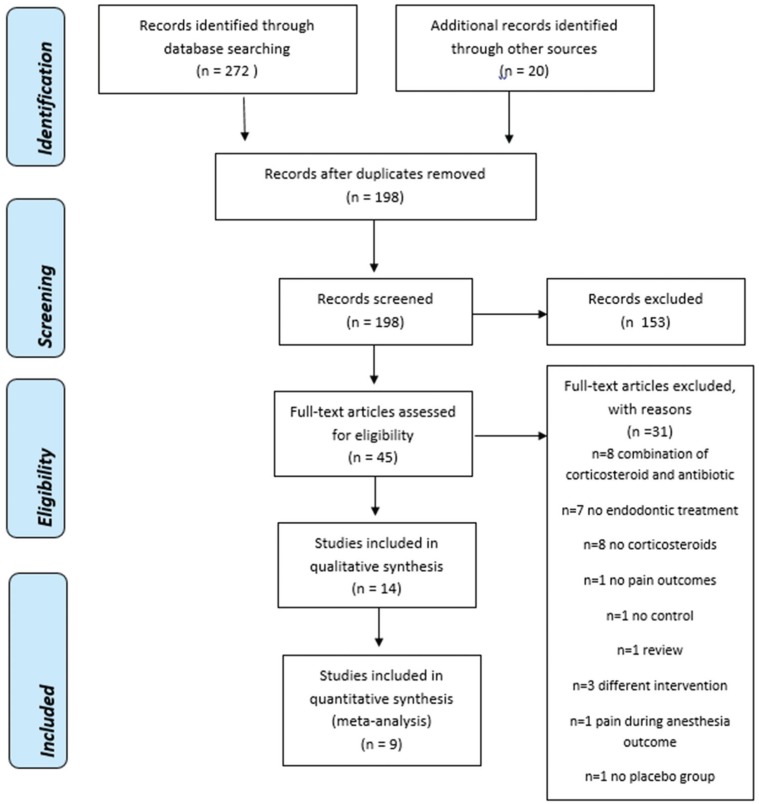
The initial search strategy yielded 178 unduplicated references plus 20 hand search references found in the bibliography section of eligible studies. Those 198 references were assessed independently by three review authors, and based on the abstracts and titles these were reduced to 40 manuscripts. Main reasons for exclusion of 153 references were that the condition was not about endodontic pain but lower back pain, or spinal pain, orthodontic pain (n = 82), the intervention was not a corticosteroid (n = 37), a duplicate reference (n = 1), no control group (n = 1), a different outcome such as efficacy of the local anesthesia (n = 10) and a review or systematic review on any topic (n = 22).
Of those 45 references, fourteen were eligible to be included in qualitative analyses [1415202122232425262728293031]. The reasons for exclusion after full-text were that the intervention was a combination of corticosteroid and antibiotic (n = 8), no endodontic treatment (n = 7), no corticosteroid (n = 8), no pain outcome (n = 1), no post- endodontic pain outcome- pain during anesthesia (n = 1) [32], no control group (n = 1), different intervention (n = 3), no placebo/no treatment group (n = 1) and a review/systematic review of the literature (n = 1). PRISMA flowchart shows a summary of our results (Fig. 1).

2. Included Studies

Study design

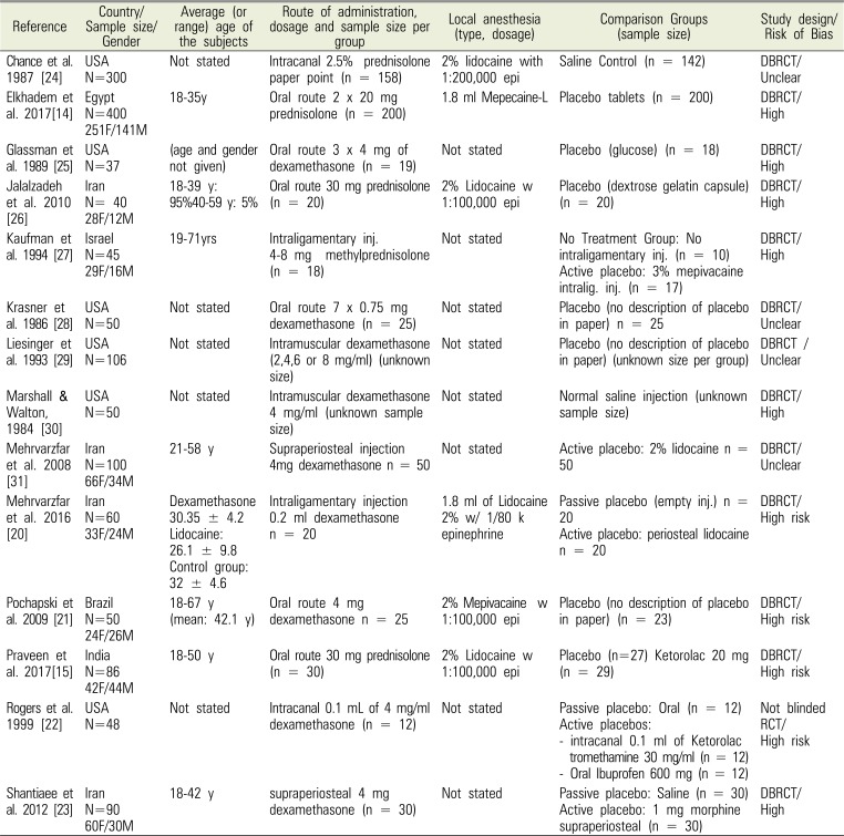
The included studies were randomized double-blinded controlled clinical trials [14152021232425262728293031] or not blinded RCTs [22] (Table 1).

Population

Patients were a mix of symptomatic, asymptomatic or both with a total of 1,462 participants. All patients needed non-surgical initial endodontic treatment. The majority of included studies were held in endodontics department of dental schools, except one which was done in a private practice [28] and two articles did not specify their clinical set up [273233].
Four studies [20232731] included only healthy adults as stated by the American Society of Anesthesiologists (ASA) categories I or II. Five studies excluded patients with allergies, systemic conditions or pregnant women [2122252630]. The gender and age of the study groups is listed in Table 1. Eight studies specified their gender ratio of their samples and F/M ratio varied from 0.9 to 2.3 [1415202123262731] with a majority of females in most of the studies. The age of the participants was between 18 to 71 (Table 1). The final number of participants in each study ranged in size from 40 [26] to 400 [14].
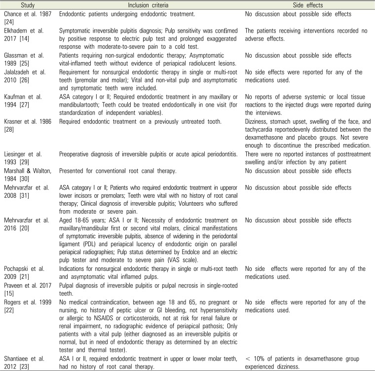
Thermal test was the most common diagnostic tool to assess the pulp vitality [1420222426], some of the studies used electrical pulp test (EPT) [142224]. In some studies, pulp vitality was an important inclusion criteria [21222531] but two studies included both vital and non-vital teeth in their intervention group [1526]. Two studies included only asymptomatic irreversible pulpitis [2125] while two studies did the opposite and only included symptomatic irreversible pulpitis [1420]. Amongst included studies, one study had the most comprehensive inclusion criteria as they included both vital and non-vital and also symptomatic and asymptomatic teeth in their study. Radiographs and diagnostic test (percussion, palpation, and biting) were used to diagnose periapical condition (symptomatic or asymptomatic apical periodontitis, acute or chronic apical abscess). The presence of pre-operative pain was specified in three studies [2931] and the absence of pain in one study [25] (Table 2). The remainder allowed both. The teeth treated varied across studies from any premolar/molar [26], upper or lower incisor or premolar [31], any molar [2023], single rooted tooth [21], to any tooth needing endodontic treatment [2122272830], and three studies made no mention of specific teeth [242529].

Intervention

Several routes of administration with various corticosteorids and dosages were administered as follows in the included studies [1415202122232425262728293031]:
  • Intracanal medication: 0.1 ml of 4 mg/ml dexamethasone or 2.5% prednisolone [2224];

  • Oral route: 1 × or 3 × 4 mg dexamethasone or 30–40 mg prednisolone or 7 × 7.5 mg dexamethasone or 4 mg dexamethasone [21252628];

  • Intraligamentary injection: 4–8 mg slow release prednisolone or 0.2 ml dexamethasone [2027];

  • Intramuscular injection: 2–4–6–8 mg/mL dexamethasone or 4 mg/ml [2930];

  • Supraperiosteal injection: 4mg dexamethasone [2331].

The intervention was a corticosteroid (dexamethasone in nine studies [202122232528293031], prednisolone in four studies [14152426], and a single study used methylprednisolone [27]. Some studies evaluated other single drug interventions besides the corticosteroid (ketorolac [22], ibuprofen [22], morphine [23]). The local anesthetic used for obtaining operative anesthesia was only mentioned in six studies: 2% lidocaine with 1:100,000 epinephrine [152126], 1:80,000 epinephrine [20] or 2% mepivacaine with 1:100,000 epi [20] and Mepecaine-L [14] (Table 1).

Comparison

Control groups included passive placebos (saline, glucose, dextrose gelatine), active placebo (2% lidocaine) in one study [31] or no treatment [27]. The only study with an active placebo group (2% lidocaine) but no passive placebo [31] was excluded of the meta-analysis to reduce heterogeneity (Table 1).

Outcomes

The method of measuring pain varied across studies and consisted of a 0–9, 0–10, 0–100 or 0–170 Visual Analog Scale or a numeric rating scale of 0–10 or 0–100. All intensity outcomes were converted to a 0–100 scale before performing a meta-analysis. The studies evaluated pain at different time points: from intraoperative; a single 24 h evaluation; and multiple evaluations at 4, 6, 12, 24, 48, 72 hours or up to 7 days. The time of drug administration was clearly specified in the studies as 30 or 60 minutes before starting the endodontic treatment, or at the time of the procedure, or immediately after the procedure, or one tab right after the endodontic procedure and one tab every 1–3 hours afterwards. The percent of patients with none or mild post-treatment pain was calculated by review authors based on data reported by the original studies in the form of graphs, tables, or in the narrative.

3. Risk of bias in included studies

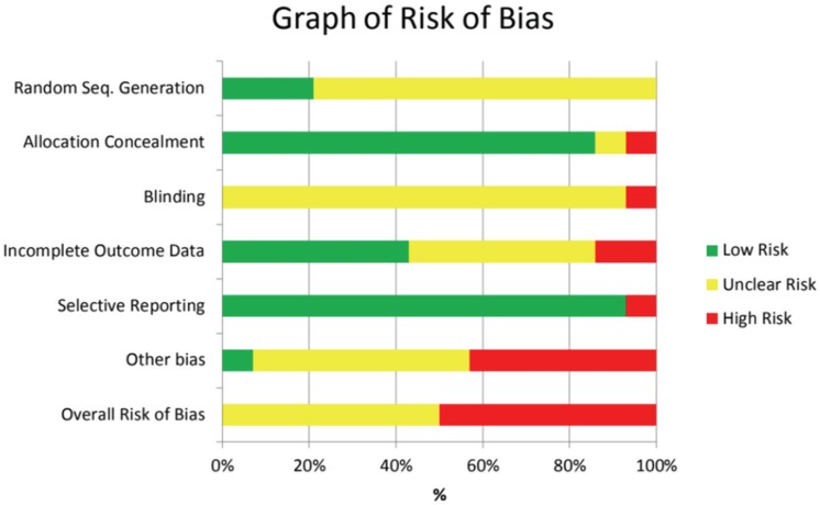
Summaries of risk of bias for each domain are shown in Table 3 (Summary of risk of bias for eligible studies) and Fig. 2 (Graph of risk of bias for eligible studies) [1415202122232425262728293031].

Random Sequence Generation

Eleven of the studies [2021222324252628293031] were classified as unclear risk of bias because they gave no description of the method or methods used to generate the random sequence in their studies. In three studies [141527] the methods of sequence generation were stated as computer-generated or using a permuted block randomization method along with an internet-based random number generator [14], and were assigned a low risk of bias in this area.

Allocation Concealment

The allocation of the participants in ten of the studies [141520212324252628293031] was low risk because, they were dispensed in disposable syringes, vials, tablets with numeric coding (by second investigator not involved in study) for treatment sequence. However it was unclear in one study where the authors claimed the study was double-blinded, but no details were provided as how the allocation was concealed [27], while in one study it was considered high risk [22], where there were no details of how the treatment groups were concealed nor any blinding strategies.

Blinding

Blinding was high risk in one study as no blinding method was described [22]. Blinding was assessed as unclear risk in eleven of the studies [14152021232425262728293031]. In each of these studies the blinding of the data analyst and technique of blinding were not described which could have resulted in bias.

Incomplete outcome data

In this category, six studies [14,15,20,22,23,24] had low risk of bias since the number of drop outs were minimal, intent-to-treat analysis was provided if needed and reasons for dropping out were balanced among treatment group. Six studies [21,25,27,28,29,31] fell into the category of unclear risk due to lack of intent-to-treat analysis [21,27,29], excluded patients due to severe pain or rescue medication [25], unclear number of excluded patients due to rescue medication [31], or total number of patients enrolled and excluded was not fully disclosed [29] or the reason for missing data were not fully disclosed [27].Two studies were considered at high risk due to the high number of patient's exclusions (>20%) [26] or the unclear description of which intervention did the participants received before reporting their pain [30].

Selective reporting

In this domain, eleven articles [14152021222324252728293031] were considered as low risk since they reported all pre-specified outcomes. One article [26] is high risk. In this article, the large numbers of excluded patients due to usage rescue medications (41% in placebo group, 31% in medication group) were not fully addressed so it is considered as high risk of bias.

Other bias

We considered six studies [142425272831] with unclear risk of other bias due to non-disclosure of their source of funding or co-interventions (i.e. parachlorophenol [24]). Four studies were at high risk of other bias due to rescue medications [21222329], occlusion adjustment [26] or lidocaine [20].

Balanced groups at baseline

The studies that did not report age and gender distribution were considered as unclear risk of bias [222425262728293031]. The ones which had balanced groups at baseline for age, gender or the intensity of pre-treatment pain were considered low risk [15202123].

Overall bias

A total of 50% of the studies were assessed at unclear risk of bias and 50% at high risk (Table 3, Fig. 2).

4. Effects of interventions

Fourteen studies were included in this review, however only nine studies could be included in the meta-analyses. Liesinger et al. 1993 [29] and Marshall & Walton, 1984 [30] failed to provide the sample size of the treatment groups or any post-endodontic treatment outcome and could not be included in the meta-analyses. Rogers et al. 1999 [22] was not blinded and did not report any outcomes of interest, precluding its inclusion in the meta-analyses. Mehrvarzfar et al. 2008 [31] reported percentage of patients with none or mild pain, however the time when the interview was conducted is unclear (6,12,24 or 48 hours after treatment) and review authors decided to exclude it from the meta-analysis to decrease bias and improve the quality of the overall results. The only study with an active placebo group (2% lidocaine) but no passive placebo namely, Mehrvarzfar et al. 2008 [31] was excluded of the meta-analysis to reduce heterogeneity. Shantiaee et al. 2012 [23] also did not report the time of the measurements (4,8,24 or 48 hours after treatment) and was also excluded.

Primary outcome (post-treatment VAS pain)

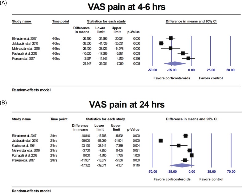
High statistically significant heterogeneity was found (Q P < .001; I2 = 96%). Post-treatment pain 4–6 hours after IANB was significantly lower by 21.147 VAS units (0–100 scale) in the corticosteroid group (95% CI = −35.034 to −7.259; P = 0.003) (Fig. 3A) and lower by 19.570 VAS units at 12 hours after endodontic treatment (95% CI = −34.772 to −4.368; P = 0.012, Forest plot not shown). However, there was no significant difference in VAS pain 24 hours after injection (Difference in means = −17.382; 95% CI = −39.071 to 4.307; P = 0.116) (Fig. 3B).

Subgroup analysis by route of administration

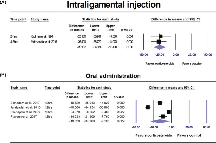
Two studies provided VAS data for intraligament administration of corticosteroids with no heterogeneity (Q P = 0.750; I2 = 0%) with similar results (fixed effect model: Difference in means = −25.167; 95% CI = −34.874 to −15.460; P < 0.001) (Fig. 4A). For oral administration of corticosteroids, however statistical heterogeneity was found (Q P < 0.001; I2 = 98%) with corticosteroids reducing significantly VAS pain (scale 0–100) compared to control at 12 hours after IANB (random-effects model: Difference in means = −19.629; 95% CI = −37.069 to −2.190; P = 0.027) (Fig. 4B).

Subgroup analysis by type of corticosteroids

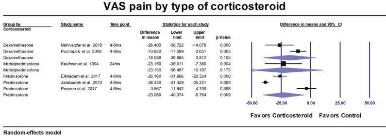
VAS pain after IANB was decreased but not significantly with dexamethasone in two studies (P = 0.105) and methylprednisone (P = 0.173), however the lack of statistical significance might be due to the small number of studies. Oral prednisolone decreased significantly postoperatively 4–6 hours after IANB with three studies (P = 0.009) (Fig. 5).

Likelihood of none or mild post-treatment pain

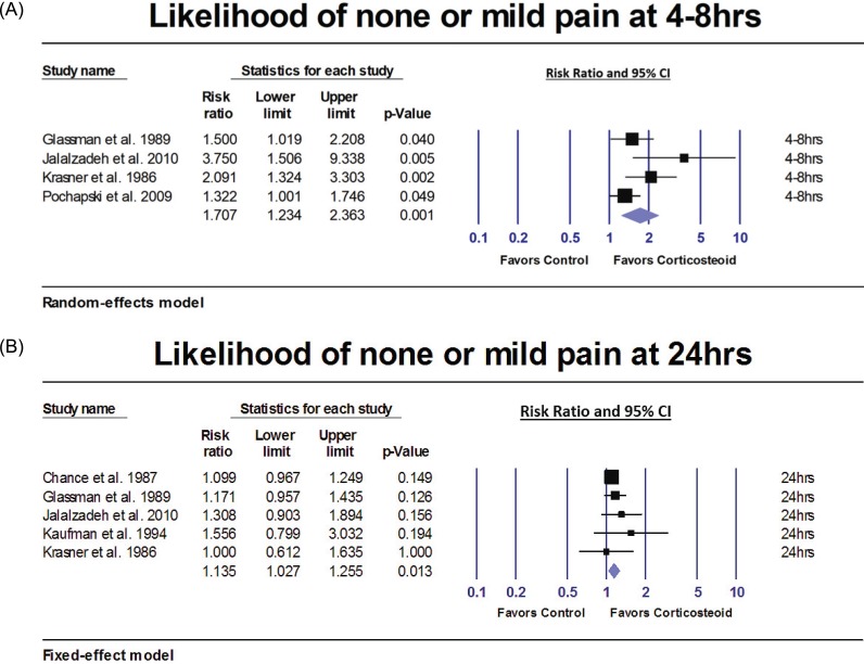
Patients who received corticosteroids prior to IANB were 70.7% more likely to have none or mild pain at 4–8 hours post-treatment (random effects model: RR = 1.707; 95% CI = 1.234 to 2.363; P = 0.001) than controls (Fig. 6A). At 24 hours post-treatment, the results were still favorable to corticosteroids with 13.5% more likely to have none or mild pain (fixed-effect model: RR = 1.135; 95% CI = 1.027 to 1.255; P = 0.013) (Fig. 6B).

Symptomatic, asymptomatic patients or both

Subgroup analyses by type of patient included in the studies found similar results. Patients in the corticosteroids groups were 50% more likely to have none or mild pain compared to controls in symptomatic patients in one study at high risk [25]; 71.3% more likely in four studies including both, symptomatic and asymptomatic patients; and 48.3% more likely in one study with only asymptomatic patients (P = 0.003) [31].

5. Adverse effects

Only one study reported a distinctive side effect of dizziness in less than 10% of its patients who took dexamethasone [23]. Very few side effects were reported by the subjects in another study, and these were evenly distributed between the dexamethasone and placebo groups. These effects were dizziness, stomach upset, swelling of the face, and tachycardia. This might not be related to the corticosteroid as it happened in both groups [28] (Table 2).

Summary of the evidence and quality of the findings (GRADE)

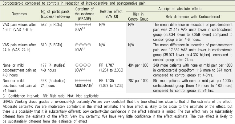
The quality of the evidence was low for all outcomes except one due to unclear/high risk of bias and statistically significant heterogeneity (Q p-value < .10 and I2 larger than 50%); low evidence grading indicates that further research is very likely to have an important impact on our confidence in the estimate of effect and it is likely to change the estimate. One meta-analysis – none or mild pain at 24 hours – did not suffer of statistical heterogeneity and was assessed at moderate quality of evidence (Table 4).

DISCUSSION

1. Main results of this review

This review included 14 RCTs with 1,462 endodontic patients. The corticosteroid drugs analyzed in this review are dexamethasone, prednisolone and methylprednisolone. Corticosteroids reduced significantly VAS pain (scale 0–100) compared to placebo by 21 units at 4–6 hours after IANB (P = 0.003) and by 19.6 units by 12 hours (P = 0.012). However, the difference was not significant after 24 hours (P = 0.116). According to Kelly 2001[34] the ‘minimum clinically significant difference’ in VAS pain is 12 units (95% CI = 9 to 15 units), so the improvements of 21 units at 4–6 hours compared to placebo (95% CI = −35.034 to −7.259; P = 0.003) on a scale 0–100 shown in this review, may be clinically significant. Based on our secondary outcome, patients who received corticosteroids prior to IANB were 70.7% more likely to have none or mild pain 4–8 hours after IANB (P = 0.001) and 13.5% more likely 24 hours after IANB (P = 0.013) than patients in the control group. A similar pattern of decreased efficiency after 24 hours appears in these results.

2. Heterogeneity of the review

The vast clinical heterogeneity amongst the 1,462 endodontic patients and interventions makes it difficult to compare the outcomes of the studies. The subjects were a mix of irreversible pulpitis and pulp necrosis, vital inflamed pulps and non-vital teeth. Inclusion and exclusion factors differed from study to study. Endodontic procedures did not follow a standard protocol. Most studies did not specify the type, dosage and technique used to gain local anesthesia. Corticosteroid intervention also varied (dexamethasone, prednisolone, methylprednisolone) as well as its concentration, route of delivery, dosage, and follow-up time (Table 1). Glucocorticoids are similar in their molecular structures and clinical action. Their main difference is in potency, duration and mineralocorticoid activity. The synthetic glucocorticoids utilized here had minimal mineralocorticoid activity and could be interchangeable. Determination of the therapeutic range could not be made as weight of patients was not noted. Both prednisolone and methylprednisolone have a half life of 60 minutes, while dexamethasone has a long half-life of 300 minutes. Based upon the VAS pain at 4–6 hours, we potentially find prednisolone and dexamethasone have similar actions, while at longer time interval the effects of the intermediate corticosteroid has its effects waning . However, further studies on this are needed.
Half-life elimination of oral dexamethasone in adults is 4 ± 0.9 hours with time to peak in serum about 1–2 hours [35]. For methylprednisolone, the half-life elimination is 2.5 ± 1.2 hours with time to peak in serum around 2.1 ± 0.7 hours [35]. In regards to oral prednisolone, half-life elimination is 2–4 hours and time to peak in serum is 1–2 hours [35]. It is important to consider the pharmacokinetics of corticosteroid medications in their efficacy of reducing post-operative pain. [35] The medications that were mostly used in our included studies have 2–4 hours of half-life elimination. It is predictable that they are not going to be as effective beyond the 4 hours point. Our meta-analysis results confirm the fact that these medications efficacy is reduced after initial 4–6 hours period.
Delivery of intervention varied between intracanal, intraligamentary, intramuscular and supra-periosteal, infiltration, oral or intracanal, however only oral and intraligamental routes were present in the meta-analyses and could be analyzed; inconclusive results for other routes. VAS pain after IANB was significantly decreased with corticosteroids delivered via intraligamental injection (P < 0.001) as well as by oral administration (P = 0.027). Insufficient data precluded analysis for other routes of administration (intracanal medication, intraligamentary, intramuscular or supraperiosteal injections). Although the time of drug administration was clearly specified in each study as either before, at the time of the procedure, right after (with or without continuing taking the medication), it is unclear when was the postoperative follow up time reference (i.e. just after local anesthetic was delivered or after surgery), which adds to the heterogeneity of the outcome data and difficulty in results comparison.

3. Agreements and disagreements with other studies or reviews

In the Compendium of Continuing Education Dental, Patten et al. 1992 reported that short term use of dexamethasone enables the clinician to take advantage of its anti-inflammatory properties and thus, dexamethasone can be an effective adjunct for postoperative control of dental pain [36]. Mohammadi [37] in the International Dental Journal, 2009 concluded that systemic steroids are highly effective in those patients who present for treatment in moderate/severe pain. Similar findings were obtained where supraperiosteal injection [31] and intraligamentary [27] injection significantly decreased postoperative endodontic pain. However, the author believes similar to our findings that it is efficacious as an adjunct to endodontic treatment, for the reduction of endodontic postoperative pain. Combining analgesics with different mechanisms or sites of action should lead to improved analgesia. Further, it can potentially reduce inflammation and provide the benefit of treating pain through different cellular pathways. A 2016 systematic review by Iranmanesh et al. [38] on the topic of pain relief following root canal treatment revealed a heterogeneity in methods and materials among the eligible studies making it impossible to perform a meta-analysis. A recent systematic review and meta-analysis by Nogueira et al. [16] studied the use of dexamethasone for controlling pain in cases of symptomatic irreversible pulpitis. In that review, only patients diagnosed with symptomatic irreversible pulpitis were included. Five RCTs [2021222531] were included in that meta-analysis. The likelihood of postoperative pain at 8 hrs, 12 hrs, and 24 hrs was compared between dexamethasone groups and other pain medications. In that review, all meta-analyses revealed a statistically significant difference favorable to the dexamethasone groups at 8 hrs (RR = 1.97, P < 0.05), 12 hrs (RR = 2.54, P < 0.05), and 24 hrs (RR = 2.58, P < 0.05).

4. Overall completeness, applicability and quality of the evidence

Four popular electronic databases (MEDLINE through PubMed, the Cochrane Library, EMBASE and Web of Science) were searched up through May 21st, 2018, for eligible studies. Authors cross-referenced and searched all included studies, literature reviews, and systematic reviews for possible missed references. Reviews, animal studies, conference proceeding abstracts, studies that were not randomized clinical trials, editorials, case reports, case series, and open-label studies were excluded by design. The results of this study could be applicable to people aged 19 years to 71 years, of both sexes, on a tooth previously untreated endodontically in need of endodontic treatment. The conclusions of this review do not apply to women who are pregnant or nursing or to children or those with endocrine disease, periodontal disease, infectious disease, or systemic disease contra-indicating endodontic therapy and any allergies to interventional medication. This systematic review included single or double-blinded RCTs as well as one not blinded study. Both, the single blinded study and not blinded were excluded of the meta-analyses to decrease bias. Thereby, the overall strength of the evidence (according to the GRADE system [17]) was low owing to unclear/high risk of bias, and statistical heterogeneity or moderate for only one outcome. The authors recommend well designed randomized, controlled, double masked trials to provide appropriate guidelines to clinicians.

5. Implications for research and clinical practice

In acute postoperative pain, various mechanisms are involved including deregulation of the inflammatory process, pain amplification and affected central inhibitory control [39]. Further central sensitization and hyperalgesia occurs due to continued pathological endodontic stimuli. Wind-up pain hyperalgesia can be an important factor during endodontic therapy due to these repeated stimulus frequencies resulting in enhancement of host cellular responses in magnitude and duration [40]. This complexity of endodontic pain, may require a multimodal approach with corticosteroids and local anesthesia to obtain superior reduction of VAS pain. Our results can help the clinician to determine the clinical significance of this multimodal therapy at various time intervals (6–8 hrs, 12 and 24 hours). In future studies corticosteroids could be considered as an adjunct for patients who are on prolonged use of NSAIDs and suffer from gastrointestinal disturbances or altered renal and hepatic function, as well as patients who have a low pain threshold. Side effects of this approach were low to non-existent. Multimodal combinations would have greater benefits by combining different mechanisms or sites of action leading to improved pain control.
This paper shows that perioperative glucocorticoids may be useful for postoperative pain reduction and can be used in clinical practice. However, a question may arise whether a second dose on postoperative day 1 can further reduce postoperative pain. Further studies are needed. Dentists are reluctant concerning the use of regular perioperative use of corticosteroids as well as for endodontic flare-ups. Overall, a multimodal analgesic approach could be used for preventing and controlling postoperative pain and provides the benefit of treating pain through different cellular pathways. One of the issues with these studies is that treatment of pain needs to be more patient-specific. Patients are not equal in pain perception due to genetic or acquired factors, resulting in them being in a high or low risk category to pain. Pain perception varies due to genetic or acquired reasons, and none of the studies or reviews took this into account. Further studies are needed to evaluate the efficacy of corticosteroids in patients with a low/high pain threshold, as well as the evaluation of secondary hyperalgesia. How many patients were completely normal after weeks or months post-endodontic therapy or did they have residual pain or hyperalgesia? Recommendation can be made to conduct further larger studies on various well-defined categories of pulp involvement patients. Further rehabilitation parameters for residual pain or hyperalgesia were not discussed either and should be looked into.

CONCLUSIONS

Our meta-analysis shows evidence of significant impact of using corticosteroid on postoperative pain reduction at 4–6 hours and 12 hours following endodontic treatment on a tooth. However, these studies could not demonstrate any remarkable effect of reducing postoperative pain at 24 hours period. We also can point out the corticosteroid effect on increasing the success rate of IANB injection, but we have to be cautious with our conclusions due to statistical heterogeneity in some analyses and unclear/high risk of bias. It is worth noting that the most common form of corticosteroid used in our included studies was oral administration of dexamethasone. Because of high risk of bias and great heterogeneity of clinical data, the authors call for more quality double blinded studies in the future to further shining light on efficacy of corticosteroid usage for pain reduction in endodontic treatment.

ACKNOWLEDGEMENTS

The authors have no funding and no conflicts of interest.

References

1. Merskey H, Bogduk N. Classification of Chronic Pain. 2nd ed. Seattle: International Association for the Study of Pain Press;1994.
2. Arora M, Sangwan P, Tewari S, Duhan J. Effect of maintaining apical patency on endodontic pain in posterior teeth with pulp necrosis and apical periodontitis: A randomized controlled trial. Int Endod J. 2016; 49:317–324. PMID: 25866134.
crossref
3. Sathorn P, Parashos P, Messer H. The prevalence of postoperative pain and flare-up in single- and multiple-visit endodontic treatment: a systematic review. Int Endod J. 2008; 41:91–99. PMID: 17956561.
crossref
4. Zuckerman O, Metzger Z, Sela G, Lin S. [“Flare-up” during endodontic treatment--etiology and management ]. Refuat Hapeh Vehashinayim. 1993; 24:19–26.
5. Rosenberg PA. Clinical strategies for managing endodontic pain. Endod Top. 2002; 3:78–92.
crossref
6. Haas DA. Local and systemic therapeutics for the control of endodontic pain. Alpha Omegan. 1997; 90:73–76. PMID: 10634107.
7. Stewart GG. Antihistamines and corticosteroids in the reduction of postoperative sequelae following endodontic surgery. Oral Surg Oral Med Oral Pathol. 1956; 9:216–220. PMID: 13297381.
8. Alexander RE, Throndson RR. A review of perioperative corticosteroid use in dentoalveolar surgery. Oral Surg Oral Med Oral Pathol Oral Radiol Endod. 2000; 90:406–415. PMID: 11027375.
crossref
9. Kidd BL, Urban LA. Mechanisms of inflammatory pai. Br J Anaesth. 2001; 87:3–11. PMID: 11460811.
10. Ingawale DK, Mandlik SK, Patel SS. An emphasis on molecular mechanisms of anti-inflammatory effects and glucocorticoid resistance. J Complement Integr Med. 2015; 12:1–13. PMID: 25503867.
crossref
11. Newton R. Anti-inflammatory glucocorticoids: Changing concepts. Eur J Pharmacol. 2014; 724:231–236. PMID: 23747654.
crossref
12. Ricciotti E, Fitzgerald GA. Prostaglandins and inflammation. Arterioscler Thromb Vasc Biol. 2011; 31:986–1000. PMID: 21508345.
13. Becker DE. Basic and clinical pharmacology of autonomic drugs. Anesth Prog. 2012; 59:159–168. PMID: 23241039.
crossref
14. Elkhadem A, Ezzat K, Ramadan M, Abdelghaffar S, Khamis D, Hassan A, et al. The effect of preoperative oral administration of prednisolone on postoperative pain in patients with symptomatic irreversible pulpitis: A single-centre randomized controlled trial. Int Endod J. 2018; 51:e189–e196. PMID: 28560802.
crossref
15. Praveen R, Thakur S, Kirthiga M. Comparative Evaluation of Premedication with Ketorolac and Prednisolone on Postendodontic Pain: A Double-blind Randomized Controlled Trial. J Endod. 2017; 43:667–673. PMID: 28320541.
crossref
16. Nogueira BML, Silva LG, Mesquita CRM, Menezes SAF, Menezes TOA, Faria AGM, et al. Is the Use of Dexamethasone Effective in Controlling Pain Associated with Symptomatic Irreversible Pulpitis? A Systematic Review. J Endod. 2018; 44:703–710. PMID: 29571913.
crossref
17. Higgins J, Green S, editors. Cochrane Handbook for Systematic Reviews of Interventions Version 5.1.0. updated March 2011.
18. Cochran W. The combination of estimates from different experiments. Biometrics. 1954; 10:101–129.
crossref
19. Higgins J, Thompson S. Quantifying heterogeneity in a meta-analysis. Stat Med. 2002; 21:1539–1558. PMID: 12111919.
crossref
20. Mehrvarzfar P, Esnashari E, Salmanzadeh R, Fazlyab M, Fazlyab M. Effect of dexamethasone intraligamentary injection on post-endodontic pain in patients with symptomatic irreversible pulpitis: A randomized controlled clinical trial. Iran Endod J. 2016; 11:261–266. PMID: 27790253.
21. Pochapski MT, Santos FA, de Andrade ED, Sydney GB. Effect of pretreatment dexamethasone on postendodontic pain. Oral Surg Oral Med Oral Pathol Oral Radiol Endod. 2009; 108:790–795. PMID: 19748294.
crossref
22. Rogers MJ, Johnson BR, Remeikis NA, BeGole EA. Comparison of effect of intracanal use of ketorolac tromethamine and dexamethasone with oral ibuprofen on post treatment endodontic pain. J Endod. 1999; 25:381–384. PMID: 10530266.
crossref
23. Shantiaee Y, Mahjour F, Dianat O. Efficacy comparison of periapical infiltration injection of dexamethasone, morphine and placebo for postoperative endodontic pain. Int Dent J. 2012; 62:74–78. PMID: 22420475.
crossref
24. Chance K, Lin L, Shovlin FE, Skribner J. Clinical trial of intracanal corticosteroid in root canal therapy. J Endod. 1987; 13:466–468. PMID: 3328770.
crossref
25. Glassman G, Krasner P, Morse DR, Rankow H, Lang J, Furst ML. A prospective randomized double-blind trial on efficacy of dexamethasone for endodontic interappointment pain in teeth with asymptomatic inflamed pulps. Oral Surg Oral Med Oral Pathol. 1989; 67:96–100. PMID: 2643074.
crossref
26. Jalalzadeh SM, Mamavi A, Shahriari S, Santos FA, Pochapski MT. Effect of pretreatment prednisolone on postendodontic pain: A double-blind parallel-randomized clinical trial. J Endod. 2010; 36:978–981. PMID: 20478449.
crossref
27. Kaufman E, Heling I, Rotstein I, Friedman S, Sion A, Moz C, et al. Intraligamentary injection of slow-release methylprednisolone for the prevention of pain after endodontic treatment. Oral Surg Oral Med Oral Pathol. 1994; 77:651–654. PMID: 8065733.
crossref
28. Krasner P, Jackson E. Management of posttreatment endodontic pain with oral dexamethasone: a double-blind study. Oral Surg Oral Med Oral Pathol. 1986; 62:187–190. PMID: 3528979.
crossref
29. Liesinger A, Marshall FJ, Marshall JG. Effect of variable doses of dexamethasone on posttreatment endodontic pain. J Endod. 1993; 19:35–39. PMID: 8289025.
crossref
30. Marshall JG, Walton RE. The effect of intramuscular injection of steroid on posttreatment endodontic pain. J Endod. 1984; 10:584–588. PMID: 6596389.
crossref
31. Mehrvarzfar P, Shababi B, Sayyad R, Fallahdoost A, Kheradpir K. Effect of supraperiosteal injection of dexamethasone on postoperative pain. Aust Endod J. 2008; 34:25–29. PMID: 18352900.
crossref
32. Aggarwal V, Singla M, Rizvi A, Miglani S. Comparative evaluation of local infiltration of articaine, articaine plus ketorolac, and dexamethasone on anesthetic efficacy of inferior alveolar nerve block with lidocaine in patients with irreversible pulpitis. J Endod. 2011; 37:445–449. PMID: 21419287.
crossref
33. Marshall JG, Liesinger AW. Factors associated with endodontic posttreatment pain. J Endod. 1993; 19:573–575. PMID: 8151248.
crossref
34. Kelly A-M. The minimum clinically significant difference in visual analogue scale pain score does not differ with severity of pain. Emerg Med J. 2001; 18:205–207. PMID: 11354213.
crossref
35. Lexi-Comp Online for Dentistry I. Lexi-Comp Online for Dentistry, Inc.;2018.
36. Patten JR, Patten J, Hutchins MO. Adjunct use of dexamethasone in postoperative dental pain control. Compendium. 1992; 13:580. 582. 584. PMID: 1521294.
37. Mohammadi Z. Systemic and local applications of steroids in endodontics: an update review. Int Dent J. 2009; 59:297–304. PMID: 19998665.
38. Iranmanesh F, Parirokh M, Haghdoost AA, Abbott PV. Effect of Corticosteroids on Pain Relief Following Root Canal Treatment: A Systematic Review. Iran Endod J. 2017; 12:123–130. PMID: 28496516.
39. Steyaert A, De Kock M. Chronic postsurgical pain. Curr Opin Anaesthesiol. 2012; 25:584–588. PMID: 22895123.
crossref
40. Fletcher MC, Spera JF. Management of Acute Postoperative Pain after Oral Surgery. Dent Clin North Am. 2012; 56:95–111. PMID: 22117944.
crossref
41. Moher D, Liberati A, Tetzlaff J, Altman DG. Preferred reporting items for systematic reviews and meta-analyses: the PRISMA statement. PLoS Med. 2009; 6:e1000097. PMID: 19621072.
crossref
Fig. 1

PRISMA Flow Diagram [41].

jdapm-18-205-g001
Fig. 2

Summary of risk of bias of eligible RCT's.

jdapm-18-205-g002
Fig. 3

(A) Results of the meta-analyses comparing corticosteroids versus controls. VAS pain was significantly decreased (P = 0.003) at 4–6 hours after IANB. (B) Results of the meta-analyses comparing corticosteroids versus controls. VAS pain was not significantly decreased after 24 hours (P = 0.116).

jdapm-18-205-g003
Fig. 4

(A) Results of the subgroup analyses by route of administration comparing corticosteroids versus control group. VAS pain was significantly decreased with corticosteroids delivered via intraligamental injection (P < 0.001). (B) Results of the subgroup analyses by route of administration comparing corticosteroids versus control group. VAS pain was significantly decreased with corticosteroids via oral administration (P = 0.027).

jdapm-18-205-g004
Fig. 5

(A) Results of the subgroup analyses by type of corticosteroids. VAS pain after IANB was decreased but not significantly with dexamethasone (P = 0.105) and methylprednisone (P = 0.173). Oral prednisolone decreased significantly postoperatively 4–6 hours after IANB (P = 0.009).

jdapm-18-205-g005
Fig. 6

A) Results of the meta-analyses comparing corticosteroids versus control group. Patients who received corticosteroids prior to IANB were 70.7% more likely to have none or mild pain 4–8 hours after IANB (P = 0.001). (B) Results of the meta-analyses comparing corticosteroids versus control group. Patients who received corticosteroids prior to IANB were 13.5% more likely to have none or mild pain 24 hours after IANB (P = 0.013) than patients in the control group.

jdapm-18-205-g006
Table 1

Summary of eligible RCT studies
jdapm-18-205-i001
Reference Country/Sample size/Gender Average (or range) age of the subjects Route of administration, dosage and sample size per group Local anesthesia (type, dosage) Comparison Groups (sample size) Study design/Risk of Bias
Chance et al. 1987 [24] USA Not stated Intracanal 2.5% prednisolone paper point (n = 158) 2% lidocaine with 1:200,000 epi Saline Control (n = 142) DBRCT/Unclear
N=300
Elkhadem et al. 2017[14] Egypt 18–35y Oral route 2 × 20 mg prednisolone (n = 200) 1.8 ml Mepecaine-L Placebo tablets (n = 200) DBRCT/High
N=400
251F/141M
Glassman et al. 1989 [25] USA (age and gender not given) Oral route 3 × 4 mg of dexamethasone (n = 19) Not stated Placebo (glucose) (n = 18) DBRCT/High
N=37
Jalalzadeh et al. 2010 [26] Iran 18–39 y: 95% 40–59 y: 5% Oral route 30 mg prednisolone (n = 20) 2% Lidocaine w 1:100,000 epi Placebo (dextrose gelatin capsule) (n = 20) DBRCT/High
N= 40
28F/12M
Kaufman et al. 1994 [27] Israel 19–71yrs Intraligamentary inj. 4-8 mg methylprednisolone (n = 18) Not stated No Treatment Group: No intraligamentary inj. (n = 10) Active placebo: 3% mepivacaine intralig. inj. (n = 17) DBRCT/High
N=45
29F/16M
Krasner et al. 1986 [28] USA Not stated Oral route 7 × 0.75 mg dexamethasone (n = 25) Not stated Placebo (no description of placebo in paper) n = 25 DBRCT/Unclear
N=50
Liesinger et al. 1993 [29] USA Not stated Intramuscular dexamethasone (2,4,6 or 8 mg/ml) (unknown size) Not stated Placebo (no description of placebo in paper) (unknown size per group) DBRCT /Unclear
N=106
Marshall & Walton, 1984 [30] USA Not stated Intramuscular dexamethasone 4 mg/ml (unknown sample size) Not stated Normal saline injection (unknown sample size) DBRCT/High
N=50
Mehrvarzfar et al. 2008 [31] Iran 21–58 y Supraperiosteal injection 4mg dexamethasone n = 50 Not stated Active placebo: 2% lidocaine n = 50 DBRCT/Unclear
N=100
66F/34M
Mehrvarzfar et al. 2016 [20] Iran Dexamethasone 30.35 ± 4.2 Lidocaine: 26.1 ± 9.8 Control group: 32 ± 4.6 Intraligamentary injection 0.2 ml dexamethasone n = 20 1.8 ml of Lidocaine 2% w/ 1/80 k epinephrine Passive placebo (empty inj.) n = 20 DBRCT/High risk
N=60 Active placebo: periosteal lidocaine n = 20
33F/24M
Pochapski et al. 2009 [21] Brazil 18–67 y (mean: 42.1 y) Oral route 4 mg dexamethasone n = 25 2% Mepivacaine w 1:100,000 epi Placebo (no description of placebo in paper) (n = 23) DBRCT/High risk
N=50
24F/26M
Praveen et al. 2017[15] India 18–50 y Oral route 30 mg prednisolone (n = 30) 2% Lidocaine w 1:100,000 epi Placebo (n=27) Ketorolac 20 mg (n = 29) DBRCT/High risk
N=86
42F/44M
Rogers et al. 1999 [22] USA Not stated Intracanal 0.1 mL of 4 mg/ml dexamethasone (n = 12) Not stated Passive placebo: Oral (n = 12) Not blinded RCT/High risk
N=48 Active placebos:
- intracanal 0.1 ml of Ketorolac tromethamine 30 mg/ml (n = 12)
- Oral Ibuprofen 600 mg (n = 12)
Shantiaee et al. 2012 [23] Iran 18–42 y supraperiosteal 4 mg dexamethasone (n = 30) Not stated Passive placebo: Saline (n = 30) DBRCT/High
N=90 Active placebo: 1 mg morphine supraperiosteal (n = 30)
60F/30M

Legend: DBRCT = Double-blinded RCT.

Table 2

Inclusion criteria and side effects reported
jdapm-18-205-i002
Study Inclusion criteria Side effects
Chance et al. 1987 [24] Endodontic patients undergoing endodontic treatment. No discussion about possible side effects
Elkhadem et al. 2017 [14] Symptomatic irreversible pulpitis diagnosis; Pulp sensitivity was confimed by positive response to electric pulp test and prolonged exaggerated response with moderate-to-severe pain to a cold test. The patients receiving interventions recorded no adverse effects.
Glassman et al. 1989 [25] Patients requiring non-surgical endodontic therapy; Asymptomatic vital-inflamed teeth without evidence of periapical radiolucent lesions. No discussion about possible side effects
Jalalzadeh et al. 2010 [26] Requirement for nonsurgical endodontic therapy in single or multi-root teeth (premolar and molar); Vital and non-vital pulp and asymptomatic and symptomatic teeth were included. No side effects were reported for any of the medications used.
Kaufman et al. 1994 [27] ASA category I or II; Required endodontic treatment in any maxillary or mandibulartooth; Teeth could be treated endodontically in one visit (for standardization of independent variables). No reports of adverse systemic or local tissue reactions to the injected drugs were reported during the interviews.
Krasner et al. 1986 [28] Required endodontic treatment on a previously untreated tooth. Dizziness, stomach upset, swelling of the face, and tachycardia reportedevenly distributed between the dexamethasone and placebo groups. Not severe enough to discontinue the prescribed medication.
Liesinger et al. 1993 [29] Preoperative diagnosis of irreversible pulpitis or acute apical periodontitis. There were no reported instances of posttreatment swelling and/or infection by any patient
Marshall & Walton, 1984 [30] Presented for conventional root canal therapy. No discussion about possible side effects
Mehrvarzfar et al. 2008 [31] ASA category I or II; Patients who required endodontic treatment in upperor lower incisors or premolars; Teeth were vital with no history of root canal therapy; Clinical diagnosis of irreversible pulpitis; Volunteers who suffered from moderate or severe pain. No discussion about possible side effects
Mehrvarzfar et al. 2016 [20] Aged 18–65 years; ASA I or II; Necessity of endodontic treatment on maxillary/mandibular first or second vital molars, clinical manifestations of symptomatic irreversible pulpitis, absence of widening in the periodontal ligament (PDL) and periapical lucency of endodontic origin on parallel periapical radiographies; Pulp status determined by EndoIce and an electric pulp tester and moderate to severe pain (VAS scale). No discussion about possible side effects
Pochapski et al. 2009 [21] Indications for nonsurgical endodontic therapy in single or multi-root teeth and asymptomatic vital inflamed pulps. No side effects were reported for any of the medications used.
Praveen et al. 2017 [15] Pulpal diagnosis of irreversible pulpitis or pulpal necrosis in single-rooted teeth.
Rogers et al. 1999 [22] No medical contraindication, between age 18 and 65, no pregnant or nursing, no history of peptic ulcer or GI bleeding, not hypersensitivity or allergic to NSAIDS or corticosteroids, not at risk for renal failure or renal impairment, no radiographic evidence of periapical pathosis; Only patients with a vital pulp (either diagnosed as an irreversible pulpitis or normal, but in need of endodontic therapy as determined by an electric tester and thermal tester). No side effects were reported for any of the medications used.
Shantiaee et al. 2012 [23] ASA I or II, required endodontic treatment in upper or lower molar teeth, had no history of root canal therapy. < 10% of patients in dexamethasone group experienced dizziness.
Table 3

Summary of risk of bias for eligible RCT studies

jdapm-18-205-i003
Study Random Seq. Generation Allocation Concealment Blinding Incomplete Outcome Data Selective Reporting Other potential bias Overall Bias
Chance et al. 1987 [24] ? - ? - - ? ?
Elkhadem et al. 2017[14] - - ? - - ? ?
Glassman et al. 1989 [25] ? - ? ? - ? ?
Jalalzadeh et al. 2010 [26] ? - ? + + + +
Kaufman et al. 1994 [27] - ? ? ? - ? ?
Table 4

Summary of the evidence and quality of the findings (GRADE)
jdapm-18-205-i004
Corticosteroid compared to controls in reduction of intra-operative and postoperative pain
Outcomes No of participants (studies) Follow-up Certainty of the evidence (GRADE) Relative effect Anticipated absolute effects
Risk in Control Group Risk difference with Corticosteroid
VAS pain values after 4–6 h (VAS 4–6 h) 582 (5 RCTs) ⊕⊕⊖⊖
LOWa,b
N/A N/A The mean difference in reduction of post-treatment pain was 21.147 VAS units lower in corticosteroid group (35.034 lower to 7.259 lower) compared to control group after 4–6 hours.
VAS pain values after 24 h (VAS 24 h) 610 (6 RCTs) ⊕⊕⊖⊖
LOWa,b
N/A N/A The mean difference in reduction of post-treatment pain was 17.382 VAS units lower in corticosteroid group (39.071 lower to 4.307 higher) compared to control group after 24hrs.
None or mild post-treatment pain at 4–8 hours 177 (4 studies)
4–8 hours
⊕⊕⊖⊖
LOWa,b
RR 1.707 (1.234 to 2.363) 494 per 1000 349 more patients with none or mild pain per 1000 in corticosteroid group(from 116 more to 674 more) compared to control group at 4–8hrs.
None or mild post-treatment pain at 24 hours 438 (5 studies)
24 hours
⊕⊕⊕⊖
MODERATEa
RR 1.135 (1.027 to 1.255) 707 per 1000 95 more patients with none or mild pain per 1000in corticosteroid group (from 19 more to 180 more) compared to control group at 24 hrs.
CI: Confidence interval; RR: Risk ratio; N/A: Not applicable
GRADE Working Group grades of evidenceHigh certainty:We are very confident that the true effect lies close to that of the estimate of the effect; Moderate certainty: We are moderately confident in the effect estimate: The true effect is likely to be close to the estimate of the effect, but there is a possibility that it is substantially different; Low certainty:Our confidence in the effect estimate is limited: The true effect may be substantially different from the estimate of the effect; Very low certainty: We have very little confidence in the effect estimate: The true effect is likely to be substantially different from the estimate of effect

aUnclear or high risk of bias in all studies, bStatistically significant heterogeneity (P < 0.10) and I2 larger than 50%.

TOOLS
Similar articles