Journal List > Brain Neurorehabil > v.10(1) > 1094148

Lee, Moon, Kwon, and Pyun: Clinical Features of an Artery of Percheron Infarction: a Case Report

Abstract

The artery of Percheron (AOP) is an uncommon variant of the paramedian artery, a solitary trunk branching off from the posterior cerebral arteries, supplying both paramedian thalami, and also often the rostral midbrain and the anterior thalamus. The typical clinical manifestations of the AOP infarction include altered mental status, cognitive impairment, and oculomotor dysfunction. We report a rare case with AOP infarction, and the clinical characteristics and rehabilitation courses for alertness disorder, cognitive dysfunction, and other accompanied symptoms.

Highlights

  • Altered mentality, memory impairment, and gaze palsy are the typical symptom triad.

  • Rehabilitative goals were set to improve the patient's alertness, cognitive functions, and gait.

  • Alertness and cognitive functions were improved during early rehabilitation period.

INTRODUCTION

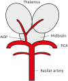
The artery of Percheron (AOP) is an uncommon variant of the paramedian artery. It is a solitary trunk branching off from either of the posterior cerebral arteries (PCA), supplying both paramedian thalami, and also often the rostral midbrain and the anterior thalamus (Fig. 1) [1]. Two large studies reported that AOP infarctions occur in a prevalence of 0.1% and 0.3% among all ischemic stroke patients, respectively [23]. Patients with AOP infarctions show a variety of clinical features, including altered mentality, memory impairment, and a vertical gaze palsy, also known as the typical symptom triad [4].
Fig. 1
Schematic diagram representing the AOP arising as a single trunk from the right PCA, supplying both thalami and often the rostral midbrain.
AOP, artery of Percheron; PCA, posterior cerebral artery.
bn-10-e2-g001
Although, there was previous case reports on AOP infarctions, none of them focused on the rehabilitative courses. In this case report, we aimed to describe a rare case of an AOP infarction patient presenting typical symptoms including altered mentality, cognitive dysfunction, and gaze palsy.

CASE REPORT

A 59-year old female patient admitted to a hospital to evaluate her dizziness and an itching sense. Peripheral origins of her dizziness were excluded through otolaryngologic evaluations. Initial magnetic resonance (MR) images of the brain showed no acute lesion or any abnormalities, but the patient's mental state drifted from alert to drowsy. Hypersomnolence persisted and she was referred to our hospital 1 week after the onset. She was regularly taking medication for her hypertension and diabetes, and had no histories of substance abuse, head injuries, or seizure activities.
Her mental state at arriving to our emergency department was drowsy, showing a Glasgow coma scale (GCS) of 14/15 (eyes 3, verbal 5, motor 6). There were no signs of focal neurologic deficits aside from drowsiness. A brain computed tomography scan did not show any abnormal findings. Laboratory tests were also within normal limits. An electroencephalogram suggested mild diffuse cerebral dysfunctions, but did not show any epileptiform discharges.
Diffusion-weighted MR imaging showed an acute infarction of both paramedian thalami and the left midbrain on the next day, suggesting an AOP infarction. MR angiography did not reveal any abnormal findings in posterior circulation, including the basilar artery (Fig. 2). Transcranial Doppler ultrasonography showed normal flow patterns and velocities in the basilar and bilateral vertebral arteries.
Fig. 2
Brain diffusion MR images (B-1000 image, axial view) showing infarctions (arrows) of (A) the bilateral thalami and (B) the left midbrain. (C) MR angiography revealed no evidence of stenosis or occlusions of posterior circulations.
MR, magnetic resonance.
bn-10-e2-g002
At the time of transfer to the rehabilitation department, 2 weeks after onset, the patient was complaining hypersomnolence, cognitive impairments, gait disturbances, diplopia, and itching on all limbs. Her mental state was slightly drowsy, obeying to 2–3 step commands. She scored 19/30 on the Korean-version of the mini-mental state examination (K-MMSE), showing defects of time orientation and attention. Motor functions were intact on all limbs. Sensory function were intact for light touch but showed decreased pin prick responses on the left forearm and calf, and decreased proprioception on both lower limbs. She scored 14/56 on the Berg balance scale (BBS) and 61/100 on the Korean-version of the modified Barthel index (K-MBI), showing moderate dependency in gait and activities of daily living. She complained vertical binocular diplopia, and a downgaze palsy of her left eye (Fig. 3).
Fig. 3
Examination of extraocular movement of the patient showing downgaze limitations of the left eye (E). (A: Upward gaze, B: Right lateral gaze, C: Primary gaze, D: Left lateral gaze, E: Downward gaze).
bn-10-e2-g003
Computerized neurocognitive function tests (CNT®; MaxMedica, Seoul, Korea) were done, revealing abnormal performance in attention, memory and learning, and executive functioning, especially in concentration, verbal long-term memory, and executive attention (Fig. 4). The patient did not show any typical aphasic or dysarthric features, but she was diagnosed with mild cognitive communication disorder according to the results of the Paradise Korea-Western Aphasia Battery-Revised (PK-WAB-R®; Paradise-Welfare Foundation, Seoul, Korea), with the aphasia quotient scores of 89.4 (90–99 percentile) and the language quotient scores of 66.7 (60–70 percentile).
Fig. 4
Results of cognitive function tests in percentiles revealing abnormal performance in memory, concentration, verbal long-term memory, and executive attention, shown through scores of CPTs, verbal learning tests, and stroop tests (color of color word, and word of color word), respectively.
F, forward; B, backward; CPT, continuous performance test; A1, first trial of list A; A5, 5th trial of list A; D, delayed; RCFT, Rey complex figure test; C, copy; I, immediate; CPM, colored progressive matrices; CPTs, continuous performance tests.
bn-10-e2-g004
Major rehabilitative goals were set to improve the patient's alertness, cognitive functions, and gait. To improve alertness and attention, 20 mg of methylphenidate and 20 mg of nicergoline were prescribed daily. Computerized cognitive rehabilitation was conducted using RehaCom® (Hasomed, Magdeburg, Germany), and the following modules were applied twice a week for 2 weeks: Aufmerksamkeit (AUFM), for concentration and scope of attention; Wortgedächtnis (WORT), for short and medium term memory; Logisches Denken (LODE), for deductive reasoning. She showed gradual advances after 4 sessions, successfully completing level 9 of AUFM, level 7 of WORT, and level 5 of LODE, improving from initial levels of 4, 4, and 1, respectively.
The patient discharged after 17 days of hospitalization. Initial complaints of pruritus, drowsiness, and hypersomnolence somewhat improved, although some daytime somnolence remained. K-MMSE, BBS, and K-MBI scores were 29/30, 55/56, and 83/100, respectively at the day of discharge. Diplopia also showed improvements, but the downgaze palsy of her left eye persisted. Prism glasses were prescribed to alleviate the diplopia.

DISCUSSION

AOP infarction shows typical symptoms of alertness problem, memory dysfunction and vertical gaze palsy due to involvement of bilateral thalami. The thalamus is a major sensory relay station, but it also processes motor inputs, limbic inputs, and inputs associated with arousal and cognitive functions, relaying them to the cerebral cortex [5]. Nuclei serving these relay functions are divided into 5 major functional classes: 1) reticular and intralaminar nuclei for arousal and nociception; 2) sensory nuclei for all major domains; 3) effector nuclei for motor and language functions; 4) associative nuclei participating in high-level cognitive functions; and 5) limbic nuclei for mood and motivation [6].
Lesions in the 4 major thalamic vascular territories (tuberothalamic, inferolateral, paramedian, and posterior choroidal artery) may result in various thalamic syndromes [6]. The AOP supplies both paramedian thalami, and also the rostral midbrain and the anterior thalamus in certain cases. These areas play important roles in maintaining consciousness and memory functioning, thus, patient affected by AOP occlusions are likely to experience memory impairments and impaired consciousness. Both anterograde and retrograde amnesia can be severe and persistent, and may accompany perseveration or confabulation [6]. In our case, decreased consciousness was the result of infarctions in both paramedian thalami and the left rostral midbrain, which then made us suspect an occlusion of the AOP, since vertebrobasilar and posterior circulations were unaffected. Like previous studies [34678910111213], our case showed severe cognitive dysfunctions, especially in attention and verbal long-term memory. Symptoms rapidly improved during early rehabilitation periods. We tried to follow up on her 6 months later, but the patient refused further evaluations. She stated during a telephone interview that there were no further memory problems. AOP infarctions generally show good functional outcomes, not hindering the independent activities of daily living [481011]. Midbrain involvement leads to relatively more unfavorable outcomes [10]. Reported long-term disability sequelae include memory deficits, impaired executive functions, behavioral problems, and oculomotor dysfunctions [4681112].
Ocular abnormalities are reported as a part of thalamic syndromes [9]. A vertical ophthalmoplegia is the most common finding among them in medial thalamic infarctions. Current knowledge states that the neural structures that control vertical gaze lie in the midbrain's reticular formation. Vertical gaze also depends on input from ascending fiber pathways from the vestibular system through the medial longitudinal fasciculus (MLF), integrating neural input into a final command. Thus, lesions affecting any of these structures can result in impaired vertical gaze. However, vertical ophthalmoplegia is also observed in patients without accompanied midbrain lesions. This might be due to disruptions of supranuclear inputs that traverse the thalamus on their way to the rostral interstitial nucleus of the MLF [14]. Management of diplopia due to supranuclear ophthalmoplegia caused by ischemic lesions consists mainly of conservative methods, such as eye patches or the use of prisms, since prognosis is good and spontaneous recovery is possible [1516].
AOP infarctions are uncommon, but its characteristic symptoms and functional impairment need comprehensive and individualized rehabilitation. If an AOP infarction is diagnosed, thorough evaluations for altered mentality, cognitive dysfunction, and oculomotor disturbances are necessary. Cognitive rehabilitation programs with the concurrent use of psychostimulants and cognitive enhancers are warranted.

Notes

Funding This study was supported by National Research Foundation of Korea (NRF-2016R1A2B4009206) funded by the Korean Government.

Conflict of Interest The authors have no potential conflicts of interest to disclose.

References

1. Percheron G. The anatomy of the arterial supply of the human thalamus and its use for the interpretation of the thalamic vascular pathology. Z Neurol. 1973; 205:1–13.
2. Carrera E, Michel P, Bogousslavsky J. Anteromedian, central, and posterolateral infarcts of the thalamus: three variant types. Stroke. 2004; 35:2826–2831.
3. Kumral E, Evyapan D, Balkir K, Kutluhan S. Bilateral thalamic infarction. Clinical, etiological and MRI correlates. Acta Neurol Scand. 2001; 103:35–42.
4. Reilly M, Connolly S, Stack J, Martin EA, Hutchinson M. Bilateral paramedian thalamic infarction: a distinct but poorly recognized stroke syndrome. Q J Med. 1992; 82:63–70.
5. Blumenfeld H. Neuroanatomy through clinical cases. 2nd ed. Sunderland (MA): Sinauer Associates;2010.
6. Schmahmann JD. Vascular syndromes of the thalamus. Stroke. 2003; 34:2264–2278.
7. Castaigne P, Lhermitte F, Buge A, Escourolle R, Hauw JJ, Lyon-Caen O. Paramedian thalamic and midbrain infarct: clinical and neuropathological study. Ann Neurol. 1981; 10:127–148.
8. Gentilini M, De Renzi E, Crisi G. Bilateral paramedian thalamic artery infarcts: report of eight cases. J Neurol Neurosurg Psychiatry. 1987; 50:900–909.
9. Bogousslavsky J, Regli F, Uske A. Thalamic infarcts: clinical syndromes, etiology, and prognosis. Neurology. 1988; 38:837–848.
10. Arauz A, Patiño-Rodríguez HM, Vargas-González JC, Arguelles-Morales N, Silos H, Ruiz-Franco A, et al. Clinical spectrum of artery of percheron infarct: clinical-radiological correlations. J Stroke Cerebrovasc Dis. 2014; 23:1083–1088.
11. Adamczyk P, Mack WJ. The artery of percheron and etiologies of bilateral thalamic stroke. World Neurosurg. 2014; 81:80–82.
12. Jodar M, Martos P, Fernández S, Canovas D, Rovira A. Neuropsychological profile of bilateral paramedian infarctions: three cases. Neurocase. 2011; 17:345–352.
13. Almamun M, Suman A, Arshad S, Kumar SJ. A case of midbrain and thalamic infarction involving artery of percheron. J Clin Med. 2015; 4:369–374.
14. Clark JM, Albers GW. Vertical gaze palsies from medial thalamic infarctions without midbrain involvement. Stroke. 1995; 26:1467–1470.
15. Barnes MP, Dobkin BH, Bogousslavsky J. Recovery after stroke. Cambridge: Cambridge University Press;2005.
16. Kim SJ, Yeom MI, Lee SU. A case of double depressor palsy followed by pursuit deficit due to sequential infarction in bilateral thalamus and right medial superior temporal area. Int Ophthalmol. Forthcoming. 2016.
TOOLS
ORCID iDs

Ahry Lee
https://orcid.org/http://orcid.org/0000-0003-1947-7602

Hyun Im Moon
https://orcid.org/http://orcid.org/0000-0003-3430-1824

Hee-Kyu Kwon
https://orcid.org/http://orcid.org/0000-0002-6230-2907

Sung-Bom Pyun
https://orcid.org/http://orcid.org/0000-0002-3471-9692

Similar articles