

 PDF
PDF ePub
ePub Citation
 Print
Print


Introduction
Paranasal sinuses are air cavities that exist in some facial bones. These vary in shape and size, and drain to the nasal cavity either directly or indirectly.12 The maxillary sinuses are the largest of the paranasal sinuses, normally segmented by septa, and are located inside of the maxillary bones.34 These sinuses can present anatomical variations, 5 extending to the anterior region of the maxilla, maxillary tuberosity, hard palate, zygomatic bone, orbit, and alveolar ridge. The extensions to the alveolar region occur in more than 80% of patients.3
In this sense, the root apices of maxillary posterior teeth may well present a close relationship with the sinus floor. The knowledge of this anatomical relationship is essential when diagnosing changes in the sinus caused by lesions of odontogenic origin or association, surgical planning, intrusion of the maxillary sinus root, fracture of the bone plate with oral sinus communication, recognition of the pathway of dental infections, and planning of orthodontic treatment.146 When there is a projection of the root into the maxillary sinus, the maxillary sinus floor deviates from its linear and horizontal path in order to bypass the dental root of the posterior teeth. This change is referred to as an 'alveolar dome' in this study.
Maxillary sinusitis is a disease that has a significant impact on a patient's health, which may include facial pain and pressure, reduction or loss of one's sense of smell, ear aches, pain in the maxilla region, as well as toothaches, fatigue, irritability, and nausea.78 As regards etiopathogeny, up to 30% of chronic unilateral maxillary sinusitis can be attributed to an odontogenic origin.9 Therefore, if the diagnosis of dental origin is not performed properly, this action can compromise treatment.10 The proximity of the dental roots to the maxillary sinus favors the dissemination of dental infections within the maxillary sinus, causing odontogenic sinusitis. Other complications, such as oroantral fistulae or root displacement after extraction, might occur due to a close relationship between dental roots and the maxillary sinus.11 Thus, knowing and identifying the relationship between these dental roots and the maxillary sinus is of utmost importance in determining proper diagnosis, planning, and treatment.
All previous studies have shown the anatomical relationship between the dental roots and the maxillary sinus through cone-beam computed tomography (CBCT).13111213 However, CBCT is not the primary exam considered for diagnosis due its high costs and radiation doses. Health history, clinical examination, and dental radiographs are necessary for diagnosis in new patients or in recall patients being evaluated for oral diseases.1415 In light of this, the present study aimed to define the term 'alveolar dome' and to evaluate the prevalence of alveolar domes in the maxillary pre-molar and molar regions using digital periapical radiographs.
Materials and Methods
This retrospective cross-sectional study was conducted after having received approval from the local Ethics Committee (CAE: 45194615.5.0000.5137). Eight hundred digital periapical radiographs from 200 dentate adult patients, who had all of their maxillary pre-molars and molars, were evaluated in regard to the presence of alveolar domes. Only the healthy maxillary sinus surrounding the teeth was considered.
All of the periapical radiographs were acquired by a digital system using the photostimulable phosphor (PSP) plate (Scan-X Duo, Air Techniques, New York, NY, USA). For the standard acquisition of images, a single operator took the radiographs, using the periapical technique of parallelism by means of Rinn XCP positioners (Dentsply, York, PA, USA). This study used the Kodak 2200 intraoral X-ray system (Kodak Dental Systems, Rochester, NY, USA), with an exposure factor of 60 kV, 7 mA, and an exposure time that varied according to manufacturer recommendations for each region (0.304 seconds for pre-molars and 0.356 seconds for molars).
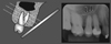
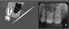
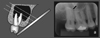
In adults, the maxillary sinuses have a pyramidal shape, extending from the root of the canine to the maxillary tuberosity, and from the floor of the orbit to the apex region of the maxillary posterior teeth. On periapical radiography, the contour of the floor of a healthy maxillary sinus is seen as a slightly curved, thin, and radiopaque line (Fig. 1).16 When pneumatization of the maxillary sinus is present, two situations may occur. In the first, pneumatization occurs in the region near the tooth root, without actually coming in contact with it. As the radiographic image is two-dimensional, the image of the maxillary sinus floor projects itself over the roots of the posterior maxillary teeth; however, it should be noted that the contour of the maxillary sinus floor remains unaltered, that is, horizontal and slightly curved (Fig. 2). In the second, the pneumatization of the real maxillary sinus comes in contact with the dental roots. Thus, the maxillary sinus floor deviates from its linear and horizontal path in order to bypass the dental root of the posterior teeth, in turn taking on a sinuous contour in the shape of a bell, with a format that is similar to the contour of the root apex, a phenomenon that we in this article term an 'alveolar dome' (Fig. 3). In this scenario, on the periapical radiograph, one can observe that the radiopaque line of the contour of the maxillary sinus floor merges with the radiopaque line of the lamina dura that bypasses the dental apex, as if both were a single sinuous and radiopaque line in close contact with the root apex.
All of the images were evaluated by 2 dentists, specialists in dental radiology and diagnostic imaging, after having been duly trained and calibrated. The interpretation of the digital images was performed directly with Kodak Dental Imaging software (Kodak Dental Systems, Rochester, NY, USA), allowing the use of all available resources. This study used a computer that contained a GeForce 9500 GT graphics card (Nvidia Corporation, Santa Clara, CA, USA) and an LED LG Flatron E2241 monitor (LG Electronics, Greater Noida, Uttar Pradesh, India) with a resolution of 1920×1080 pixels, together with brightness and contrast levels of the monitor set to their pre-defined configurations.
BioEstat 5.0 software (Instituto de Desenvolvimento Sustentável Mamirauá, Belém, Pará, Brazil) was used to compare the prevalence of alveolar domes among the maxillary teeth and, considering the molars, to compare the prevalence of alveolar domes among the different roots of the same tooth. The χ2 test was applied with a significance level of 5%.
Results
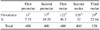
The prevalence of alveolar domes was evaluated in 400 first pre-molars, 400 second pre-molars, 400 first molars, 400 second molars, and 128 third molars. The results demonstrated that the prevalence of alveolar domes identified in the first pre-molars was 7.75% (31/400), which is statistically significantly lower when compared to the other maxillary posterior teeth (19.25% for second pre-molar [77/400], 30.5% for first molar [122/400], 32% for second molar [128/400]), and 22.66% for third molars (29/128) (p<0.05) (Table 1). There was also no statistically significant difference in the prevalence of alveolar domes between the first and second maxillary molars, and between the second pre-molar and third molars. However, for the second pre-molars and third molars, the prevalence of alveolar domes was statistically lower when compared to the first and second molars and statistically higher when compared to the first pre-molars (p<0.05).
In the evaluation of the presence of alveolar domes among the roots of the first molars, it was observed that the palatal (P) root presented a lower prevalence of alveolar domes (11.25%, 45/400) when compared to the distobuccal (DB) (28.25%, 113/400) and the mesiobuccal (MB) (29.75%, 119/400) roots (p<0.05). The buccal roots presented no statistically significant differences among them (Table 2).
The same characteristics found for the roots of the first molars were also identified for the maxillary second molars. In other words, no statistically significant difference was observed in the prevalence of alveolar domes among the buccal roots (30.25% for MB root [121/400], and 29% for DB root [116/400]), which present a higher prevalence of alveolar domes when compared to the 13.5% for the P root (54/400) (p<0.05) (Table 3).
Discussion
In this study, the prevalence of alveolar domes in maxillary sinus was examined using periapical radiographs.
Due to the anatomical proximity between the maxillary sinus and the root apices of the posterior teeth, various cases of maxillary sinusitis are of odontogenic origin or association with periapical and periodontal lesions, as well as tooth extractions.917 In addition, the endodontic treatment of pre-molars and molars can result in accidents, such as oral sinus communication, allowing for the displacement of infected tissues to the inner portion of the maxillary sinus, which can cause acute or chronic forms of sinusopathy.18
The distance between the maxillary sinus and the roots of maxillary posterior teeth were analyzed by Kilic et al.3 through 92 computed tomography (CT) images. These authors maintained that the roots of the first pre-molars had less contact with the maxillary sinus, whereas the buccal roots of the second molars had more contact but presented no statistically significant difference between them. These results are quite similar to those from the present study, which observed that the first pre-molars present a lower prevalence of alveolar domes, while the buccal roots, as compared to the palatal root, of the maxillary first and second molars present a greater prevalence of alveolar domes. This affirmation can be explained by the anatomy of the maxillary sinus, which shows a tendency towards a reduction in volume in the medial and posterior directions.19
One study performed by Pagin et al.,2 conducted using CT images, verified that the root apices protruded into the maxillary sinus in 21.1% of the first pre-molars, 22.2% of the second pre-molars, 20.3% of the first molars, 25% of the second molars, and 11.1% of the third molars. In the present study, it was observed that the root apices protruded into the maxillary sinus in 7.75% of the first pre-molars, 19.25% of the second pre-molars, 30% of the first molars, 32% of the second molars, and 22.66% of the third molars. Upon comparing the two studies, a greater difference was found in the prevalence of the alveolar domes of the first pre-molars and third molars; however, this difference may well be related to the different types of exams used in each study.
As regards the roots of the maxillary first and second molars, many studies have demonstrated that the MB roots of these molars were most frequently associated with alveolar domes when compared to the DB and P roots, and that the P roots contained the lowest prevalence of alveolar domes.121320 The present study also observed a lower prevalence of alveolar domes in the P roots, when compared to the buccal roots (p<0.05). However, no statistically significant difference was observed between the MB and DB roots (p>0.05). This divergence can be explained by the sensitivity of the diagnostic method, given that the present study was conducted with digital periapical radiographs, while the other studies were conducted using CT exams. The same was not true for the first pre-molar and the P root of the first and second molars, as they presented results that were similar to prior studies, that is, a lower prevalence of alveolar domes.81320
No previous study using periapical radiographs has evaluated the anatomical relationship between the apices of the maxillary posterior teeth and the maxillary sinus floor. In this study, the results of the prevalence of alveolar domes, using two-dimensional periapical radiographs as an evaluation method, were similar to those found in works that used three-dimensional CT exams.681319202122 However, periapical radiographs have the advantage of being an imaging method that is more commonly used by dentists, due to their cost, accessibility, and lower radiation dose.21 Once the periapical radiograph has identified an alveolar dome, the decision to recommend a CT exam should be based on the patient's history and clinical examination. 23
In conclusion, the present study coined the term 'alveolar dome,' referring to the anatomical projection of the root into the floor of the maxillary sinus. In regard to prevalence, this study showed that the first and second molars presented a greater prevalence of alveolar domes, especially in the buccal roots, followed by the third molars and second pre-molars. The first pre-molars presented a lower prevalence of alveolar domes. Although the periapical radiograph is a two-dimensional method, the results of this study showed that periapical radiographs can provide dentists with the auxiliary information necessary to identify alveolar domes, improving diagnosis, planning, and treatment.


 XML Download