

 PDF
PDF ePub
ePub Citation
 Print
Print


REFERENCES
1. The International Consensus Group for Hepatocellular Neoplasia. Pathologic diagnosis of early hepatocellular carcinoma: a report of the international consensus group for hepatocellular neoplasia. Hepatology. 2009; 49:658–664.
2. Hytiroglou P, Park YN, Krinsky G, Theise ND. Hepatic pre-cancerous lesions and small hepatocellular carcinoma. Gastroenterol Clin N Am. 2007; 36:867–887.

3. Kojiro M, Roskams T. Early hepatocellular carcinoma and dysplastic nodules. Sem Liv Dis. 2005; 25:133–142.
4. Theise ND, Park YN, Kojiro M. Dysplastic nodules and hepatocarcinogenesis. Clin Liver Dis. 2002; 6:497–512.
5. Terminology of nodular hepatocellular lesions. International working party. Hepatology. 1995; 22:983–993.
6. Theise N, Park C, Kim YB, Chae KJ, Park YN. Apoptosis and proliferation in hepatocarcinogenesis related to cirrhosis. Cancer. 2001; 92:2733–2738.
7. Theise ND, Thung SN, Cubukcu O, Fernandez GJ, Yang CP, Park YN. Neoangiogenesis and sinusoidal "capillarization" in dysplastic nodules of the liver. Am J Surg Pathol. 1998; 22:656–662.
8. Park YN, Kojiro M, Di Tommaso L, et al. Ductular reaction is helpful in defining early stromal invasion, small hepatocellular carcinomas, and dysplastic nodules. Cancer. 2007; 109:915–923.
9. Di Tommaso L, Destro A, Seok JY, et al. The application of markers (HSP70 GPC3 and GS) in liver biopsies is useful for detection of hepatocellular carcinoma. M J Hepatol. 2009; 50:746–754.
Fig. 1.
Gross findings of explant-ed liver. The serial cut surfaces of the liver showed multiple hepatocellular carcinomas (HCC1-3), early hepatocellular carcinomas (eHCC1-7), high-grade dysplastic nodules (DNHG1-8), low-grade dysplastic nodules (DNLG), and large regenerative nodules (LRN1-2) in the background of B viral and alcoholic macronodular cirrhosis.
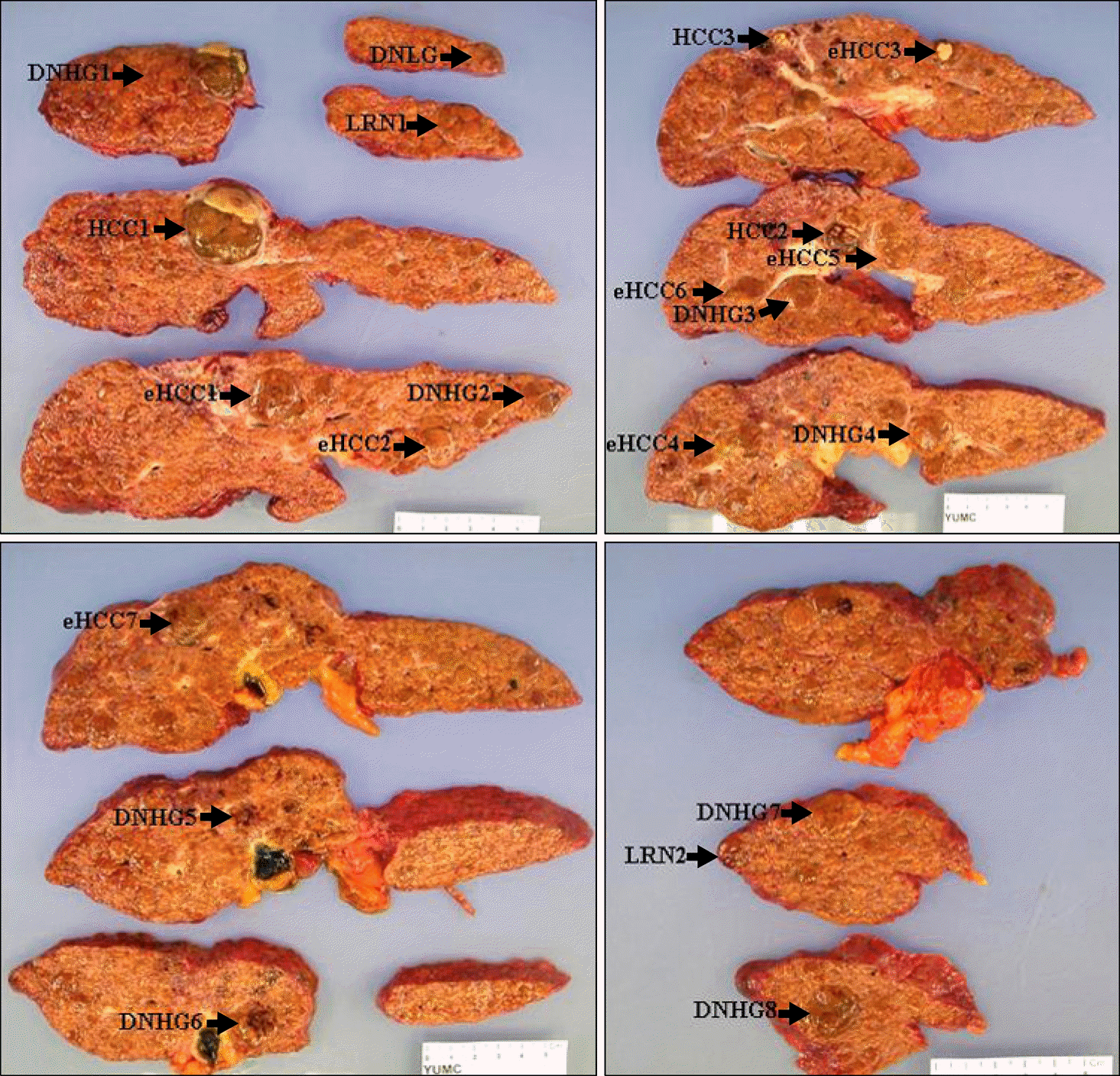
Fig. 2.
Hepatocellular carcinoma and high-grade dysplastic nodule. (A) A 2 cm sized hepatocellular carcinoma (HCC 2 in Fig. 1B, arrow heads) and a 1.5 cm sized high-grade dysplastic nodule (DNHG3 in Fig. 1B) are found grossly. (B) Microscopic finding of hepatocellular carcinoma showed moderate differentiation (H-E, ×100). (C, D) Dysplatic nodule, high grade (arrows) showed increased cellularity compared to surrounding cirrhotic nodules and small liver cell change (C: H-E, ×20; D: H-E, ×100).
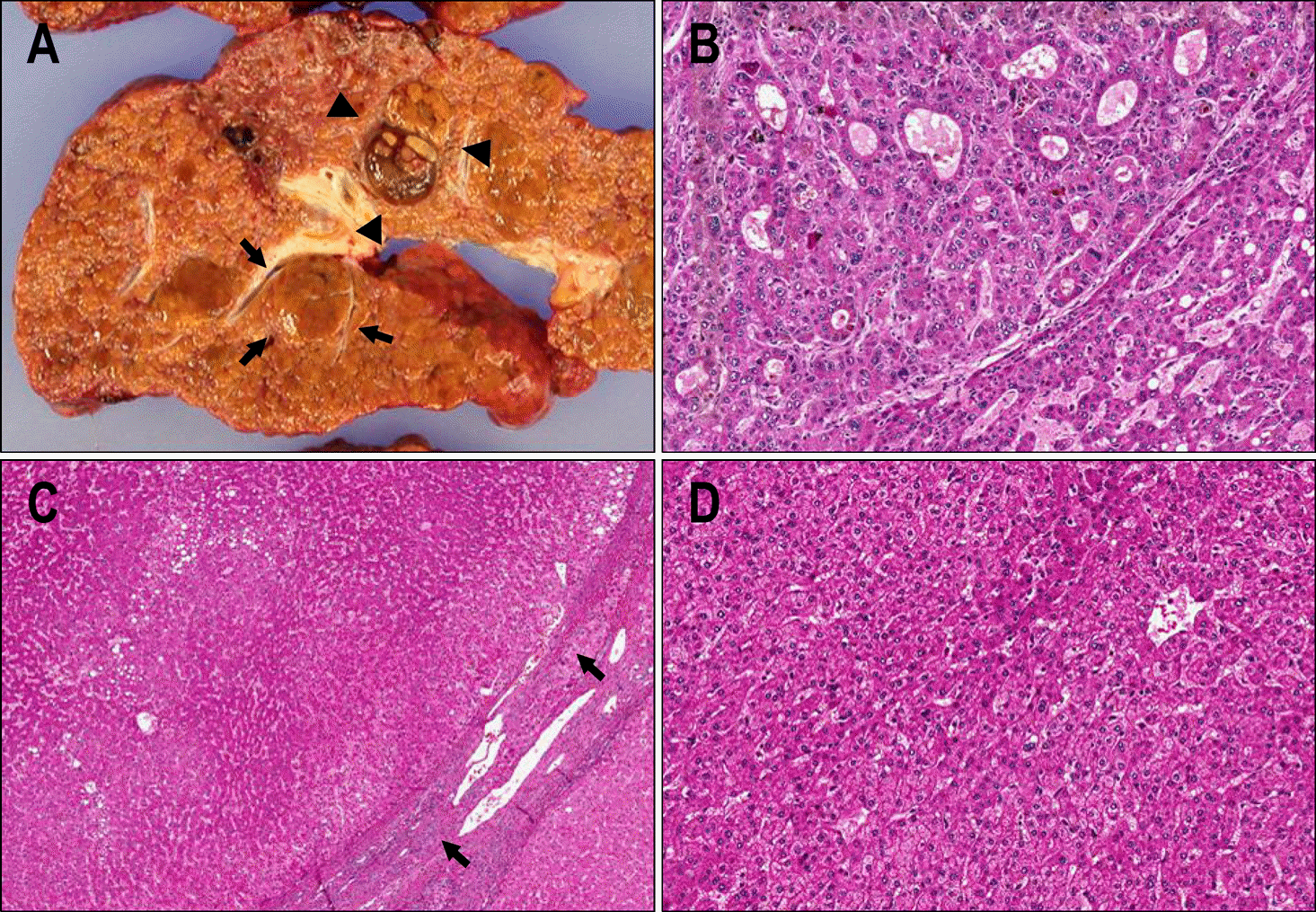
Fig. 3.
Early hepatocellular carcinoma. (A) A 2 cm sized and brown colored nodule (arrows) (eHCC1 in Fig. 1A) was noted in the cirrhotic liver. (B) On scan power, nodular lesions (arrows) show cellularity increased more than two times compared to that of cirrhotic nodule, and it is sur-rounded by fibrous septa of background cirrhosis (H-E, scan power). (C) Well differentiated hepatocellular carcinoma showed nor-motrabecular pattern, several un-paired arteries without accompanying bile duct and stromal invasion of tumor cells (arrows) (C: H-E, ×20; D: H-E, ×100).
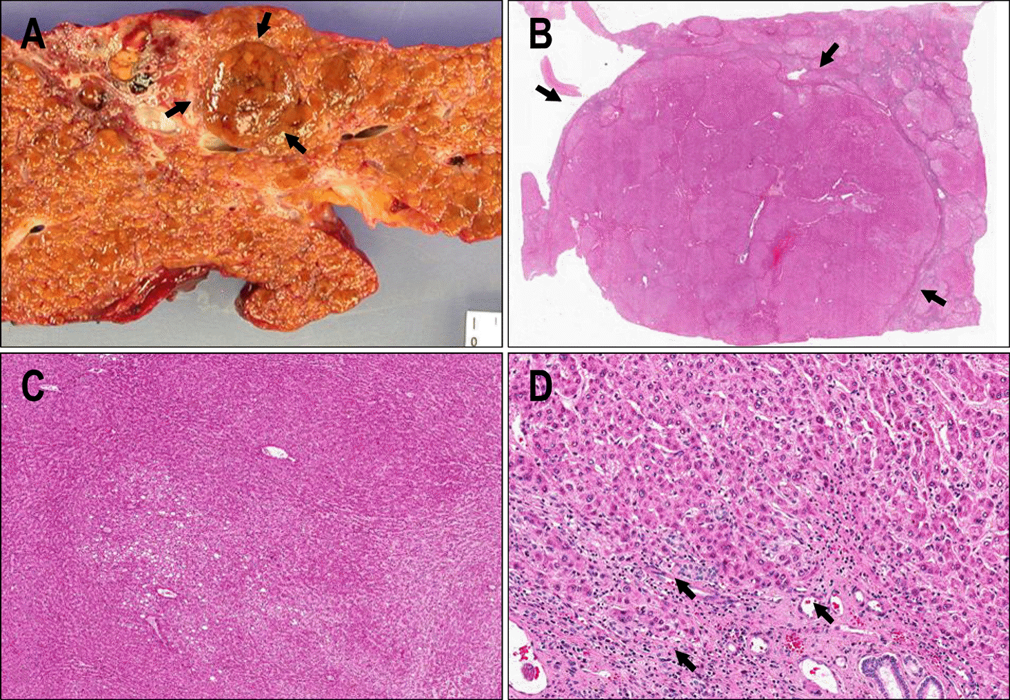
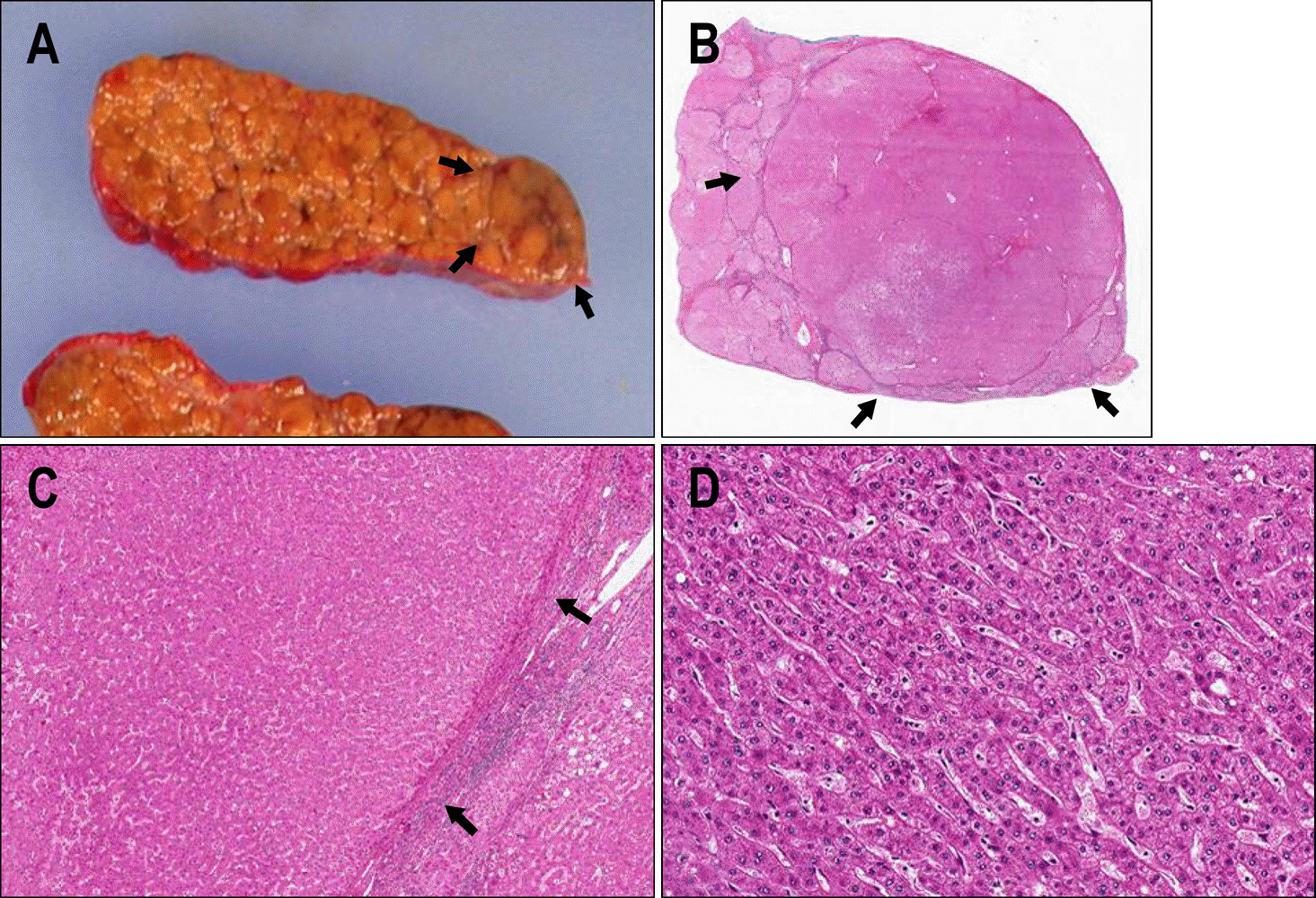
Fig. 4.
Low-grade dysplastic nodule. (A) A 1 cm sized, large nodule (DNLG in Fig. 1A) (arrows) is noted under the Glisson's ca-pule grossly. This nodule was dis-tinguished from the adjacent cirrhotic nodules due to its size, col-or, and bulging out texture. (B-D) The low grade dysplastic nodule (arrows) showed mild dysplasia and a slightly increased cellularity compared to surrounding cirrhotic nodules. Several portal tracts were found in the dysplastic nodule (B, H-E, scan power; C: H-E, ×20; D: H-E, ×100).
 XML Download