

 PDF
PDF ePub
ePub Citation
 Print
Print


INTRODUCTION
Endoplasmic reticulum (ER) plays a major role in protein synthesis, folding and processing of secretory and transmembrane proteins [12]. ER stress is caused by the accumulation of misfolded or unfolded proteins in ER due to genetic or environmental factors [3]. This process is mediated by three ER transmembrane domains: IRE1, PERK and ATF6, which induce the Unfolded Protein Response (UPR). First, translational attenuation due to PERK-dependent phosphorylation of eIF2 occurs to overcome the burden of ER [4]. Failure of this process induces UPR-related genes, primarily chaperones such as the immunoglobulin binding protein (BiP, a 78 kDa glucose control protein GRP78) and 74 kDa glucose control protein (GRP74), to prevent further accumulation of unfolded and/or misfolded proteins, with simultaneous transcriptional activation of the UPR genes such as IRE1α / XBP1 and AFT6. Activation of IRE1α by ER stress results in generation of the XBP1 spliced form by inducing the endonuclease domain of IRE1α to cleave the 26-nt intron from full-length XBP1 mRNA [5]. Persistence of the problem results in abnormal proteins in the ER being extracted, reversed to the cytoplasm, and degraded by an ER related Associated Degradation (ERAD) system consisting of ubiquitin-dependent proteasomes [6]. This stimulates the nuclear factor kappa light chain-enhancer of activated B cells (NF-κB), a transcription factor mediating immunity and anti-apoptotic responses. Irreversible cellular damage and insufficient pro-survival activity of the UPR for the retention of cellular homeostasis results in induction of apoptosis by stimulation of the CCAAT/enhancer-binding homologous protein (CHOP) and activation of the JNK (c-Jun N-terminal kinase) kinase and caspase-12 [78].
ER stress and UPR are critical to intestinal cells, and damage to UPR signaling leads to chronic inflammatory diseases such as Inflammatory Bowel Disease (IBD), Rheumatoid Bowel Syndrome (IBS), and Necrotizing Enterocolitis (NEC) [910].
Asian countries have seen a dramatic increase in IBD incidence due to westernized diet patterns [11]. IBD is a complex disease caused by several genetic and environmental factors. The cause of Crohn's Disease (CD) and Ulcerative Colitis (UC), two principal types of IBD, are intestinal inflammation and ER stress, especially in the ileum and/or colonic epithelia [1213]. Inflammatory symptoms are augmented by diet patterns such as gluten sensitivity, alterations in the gut microbiome, breach of intestinal barrier and genetic susceptibility [14]. Breach of intestinal barrier and alteration of the gut microbiome cause dysfunction of the innate immune system such as toll-like receptors [15]. Genome-Wide Association Study has confirmed 163 IBD susceptibility loci related to 300 known genes, which in turn are associated with cytokine induction, lymphocyte activation, response to bacterial infection, and ER stress [16171819]. Hence, IBD patients have increased levels of cytokines such as tumor
necrosis factor-alpha (TNFα), interferon-gamma (IFNγ), interleukin (IL) 1β, IL6 and IL17 [20]. Furthermore, due to loss of integrity of the intestinal epithelium, nutritional deficiencies (such as malabsorption, diarrhea, and intestinal blood loss) are common features of IBD patients. Therefore, dietary interventions are extremely important for these patients, which include certain vitamins and mineral replacement therapy, low fiber diets and low-FODMAP diet (the global restriction of all fermentable carbohydrates) [212223]. Recent alternative approaches use alternative medicines for IBD therapy [24]. Herbal therapy with curcumin in UC, and wormwood in CD, were explored for IBD patients [2526].
Although a native plant grown only in the Ningxia Hui Autonomous Region of north-central China, Lycium barbarum (L. barbarum) has gained considerable attention as a superfood in the western countries [27]. It has long been used as an edible and traditional medicinal plant in eastern countries such as China and Korea [28]. Fresh or dried fruits are used as food, and leaves are used as food or tea. Of the various components of LF, the L. barbarum polysaccharides (LBP) are the most well-studied. Other components include carotenoids and their related compounds, betaine, cerebroside, β-sitosterol, p-coumaric acid, and vitamins [29]. L. barbarum has been administered for the treatment of diabetes and hyperlipidemia, for reducing blood sugar and serum lipids, respectively, and is also known to exert its effects of neuroprotection and antioxidants [30313233]. However, the outcomes of LF on ER stress and oxidative stress on intestinal epithelial cells are not well known. This study was therefore undertaken to investigate the anti-inflammatory effects of LF associated to the ER stress pathway on polarized human intestinal epithelial cells.
MATERIALS AND METHODS
Materials
Dried 5 kg of LF was harvested from the Ningxia Hui Autonomous Region of north-central China; extraction was performed for 7 days by adding 70% alcohol to make 8 times of the weight. The resultant extract was filtered, concentrated in a rotary evaporator, and used in this study.
Cell culture and cell viability
Human intestinal epithelial Caco-2 cells and mouse embryonic fibroblast (MEF) cell lines (American Type Culture Collection, USA) were cultured in Dulbecco's modified Eagle's medium (DMEM, Gibco, USA) supplemented with 10% heat-inactivated fetal bovine serum (FBS, Gibco, Grand Island, NY, USA) and 1% Penicillin-Streptomycin solution (P/S, Sigma-Aldrich Co., USA) at 37℃ under humidified 5% CO2. Caco-2 polarization was performed as follows: 24-well Transwell (35024, SPL, Korea) dishes were coated with 396 µg/cm2 collagen, seeded with Caco-2 cells, and monolayers were grown for 10 days. The experiment was performed when the Transepithelial/Transendothelial Electrical Resistance (TEER) reached a value in the range of 500–600 Ωcm2. LF stock solution (100 mg/mL) was made by dissolving 100 mg of the dry crude extract in 1 mL DMSO. Cell viability was determined by the WST assay kit (EZ-cytox, Daeil lap service Co. Ltd., Seoul, Korea). Caco-2 cells were seeded in 96-well plate and incubated for 24 hours (h) under the same culture conditions previously mentioned. Cells were pretreated with various concentrations of LF (0, 12.5, 25, 50, 100 and 250 µg/mL) for 24 h followed by WST assay, according to the manufacturer's instruction. Absorbance was measured at 450 nm on a micro-plate reader (xMark™ Microplate Absorbance Spectrophotometer, Bio-Rad Inc., Hercules, California, USA) after 3 h.
RNA extraction and cDNA synthesis
Total RNA was extracted from polarized Caco-2 cells and MEF cell lines. Briefly, after removal of the cell medium, Tri-reagent (MRC Inc., Cincinnati, USA) was added to each well and the resultant lysates were harvested. Chloroform was then added, and the mixture was centrifuged at 12,000 g for 15 minutes at 4℃. The supernatant was separated, mixed with isopropanol, and centrifuged at 12,000 g at 20℃ for 8 minutes. RNA pellets were harvested, and the RNA concentration was measured using Nano Drop (Nano Drop ONE, Thermo Scientific Inc., USA), after which cDNA was synthesized using the RT-Kit (M-MLV, RNase H -, BioFACT Co., Korea).
Real-time qPCR
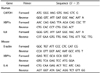
The synthesized cDNA was analyzed by the real-time PCR system using 2X Real-Time PCR Master Mix kit (Including SYBR Green, Low ROX, BioFACT Co., Korea) according to manufacturer's instruction. Amplification was performed for 45 cycles. Data analysis was performed using the Agilent AriaMX 1.0 program. The primer sequences used in the experiments are presented in the Table 1.
Reverse transcription-PCR
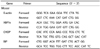
In order to measure mRNA expression of the X-box binding protein 1 spliced form (XBP1s), reverse transcription (RT)-PCR was performed according to the manufacturer's instruction (2X Taq Basic PCR Master Mix2, BioFACT Co., Korea). The primer sequences used in the experiments are presented in Table 2. The samples were loaded onto a 3% agarose gel for electrophoresis and verified under UV light (AE-9000 E-Graph, ATTO, Japan).
Paracellular permeability
Caco-2 cells polarized for 10 days were pretreated with LF extract for 24 h, followed by exposure to a cytokine cocktail (50 ng/mL TNF-α + 50 ng/mL IFN-γ + 25 ng/mL IL-1β + 10 µg/mL LPS) applied on the apical side of the chamber for an additional 24 h. Apical and basal side were then washed with HBSS (Hanks' balanced salts, Sigma-Aldrich Co., USA) supplemented with 10 mM HEPES (Sigma-Aldrich Co., USA). Fresh HBSS/HEPES was added to the basal side, and 4 kDa fluorescein isothiocyanate dextran (FITC-dextran, Sigma-Aldrich Co., Sweden) diluted with HBSS/HEPES solution to a final concentration of 1 mg/mL was added to the apical chamber and incubated for 72 h. At the indicated time point, the basal solutions were collected and the fluorescence signal was detected using a DTX 800 multimode detector (DTX800, Beckman Coulter Inc., Brea, CA, USA) at excitation wavelength of 490 nm and emission wavelength of 520 nm.
LF treatment on Zebrafish embryo
Wild type (wt) zebrafish (D. rerio) were maintained at the Chungnam National University (CNU-01027) as per the Animal Research Guidelines. Wild type zebra fish and/or eggs were provided by the Zebrafish Center for Disease Modeling (ZCDM), Korea. At 2 to 75 h post-fertilization (hpf), zebrafish embryos were isolated in 24-well plates (10 embryos/well) in 1 mL standard egg water (0.1%) supplemented with methylene blue, then they were exposed to LF (0, 12.5, 25, 50, 100, 250 µg/mL) for various times as indicated. The LF solutions were changed once every 24 h. Embryos were imaged until hatching, using the Leica microscope (DM2000, Leica co., Wetzlar, Germany) and Canon microscope (SZ2-ILST, Tokyo, Japan).
Scanning ion-conductance microscopy (SICM)
SICM images were obtained with a commercial SPM (NX- 12 system, Park Systems Corp, Suwon, Korea). This system has a Scanning Probe Microscope system fixed on top of an inverted optical microscope (Eclipse Ti, Nikon Corp., Tokyo, Japan) enabling to view the bottom side of the sample and acquire SICM images simultaneously. The SPM system has a 100 µm × 100 µm xy flat scanner, and a separate SICM head with a 15 µm z scanner. The SICM probe consists of a glass pipette filled with electrolyte, and an Ag/AgCl pipette electrode inserted into it. The SICM probe is fabricated from borosilicate capillaries (inner diameter 0.58 mm, outer diameter 1.0 mm, Warner Instruments, Hamden, CT, USA) using a CO2-laser-based micropipette puller (P-2000, Sutter Instruments, Novato, CA, USA); the inner and outer diameters of the tip of the SICM probe are approximately 80 nm and 160 nm, respectively. In the present study, the surface profiles of fixed single cell images were primarily obtained using the Approach-Retract Scanning (ARS) mode of the SICM to ensure non-destructive high resolution surface images.
Statistical analysis
Data from all experiments were analyzed using the SPSS/Windows 24.0 (SPSS Inc., Chicago, USA) program. All experiments were carried out more than 3 times. Results are expressed as mean ± SD. Student's t-test was used to obtain the mean difference between two groups, and One-way ANOVA was used for two or more groups. After ANOVA analysis, Duncan's multiple range test was applied to identify differences between groups. A P-value < 0.05 is considered statistically significant.
RESULT
In vitro and in vivo toxicity of LF extract
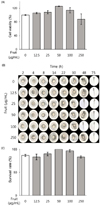
To investigate the effect of LF on cell viability, WST assay was performed on Caco-2 cells (a human colon epithelial cell line). This assay converts the dehydrogenated and water-soluble tetrazolium of living cells to a detectable aqueous soluble formazan (orange color), thereby identifying cell proliferation. No change in cell viability was observed after exposing the cells to LF extract for 24 h, suggesting that the extract is non-toxic in the range 0–250 µg/mL LF in vitro (Fig. 1A).
To examine the in vivo toxicity of LF, zebra fish embryos treated with different concentrations of LF dissolved in egg water were observed for development and documented at each time point (Fig. 1B). No deterioration of embryo development or hatching time variation were observed after exposure to LF up to 250 µg/mL. Although a shorter hatching time was observed at 25 and 100 µg/mL LF, there were no changes of development at any of the tested concentrations. The number of embryos hatched were counted and calculated for the survival rate, presented in Fig. 1C. No statistically significant difference was observed between the indicated concentrations for the survival rate, suggesting the range of LF concentration between 0 µg/mL and 250 µg/mL in vivo is not toxic. Based on these data, LF concentrations used in all subsequent experiments were determined to be less than 100 µg/mL, and were concluded to be safe.
Evaluation of paracellular permeability in polarized Caco-2 cells after LF treatment
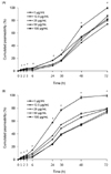
Maintaining TJ integrity in the intestine is critical for nutrient absorption, host defense, and host immunity. Alteration of intestinal TJ homeostasis is thought to induce pathogenesis of IBD as well as obesity [34]. Since polarized Caco-2 cells have similar structures and functions as intestinal cells and are useful for observation of cell permeability [35], we investigated the effect of LF on the TJ integrity in these cell types. The polarized Caco-2 monolayers were pretreated with LF for 24 h, followed by an additional 24 h incubation without (Fig. 2A) or with (Fig. 2B) cytokine cocktail (50 ng/mL TNF-α + 50 ng/mL IFN-γ + 25 ng/mL IL-1β + 10 µg/mL LPS). Subsequently, 1 mg/mL FITC-dextran was added at the apical section of the chamber, and the fluorescence intensity of the basal chamber was measured to examine the permeability of TJ at each indicated time point. We observed that FITC-dextran in the basal chamber accumulated less significantly in cells pretreated with LF than in untreated cells. Except for 100 µg/mL LF, the accumulated permeation decreased at higher concentrations in a dose-dependent manner (Fig. 2A). Cells stimulated with the cytokines represent the inflammation status in this study. These cells showed a similar pattern, wherein LF pretreatment lowered the permeability of FITC-dextran as compared to the untreated cells, even in the presence of inflammation (Fig. 2B). Taken together, these results indicate that LF has a protective effect of maintaining the TJ integrity of the polarized Caco-2 monolayer, both in the presence and absence of external inflammatory stimuli.
Expression of IL8 by LF in human intestinal epithelial cells
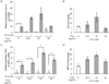
It is well known that ER stress induces a proinflammatory response [36]. To investigate the effect of LF on the expression of proinflammatory cytokines, polarized Caco-2 cells were treated with LF for 24 h, after which ER stress was induced by treatment with 3 µM Thapsigargin (TG). qRT-PCR was performed to examine the expression of representative proinflammatory cytokine IL8, followed by GAPDH normalization (Fig. 3). Compared to untreated cells, cells treated with TG showed significant induction of IL8 (left bars of Fig. 3A), suggesting that TG stimulation works as a positive control in our study. More importantly, we observed that LF itself had no effect on IL8 induction. Fig. 3B shows the fold change by TG stimulation at each concentration of LF pretreatment. A trend was observed for IL8 mRNA expression levels which showed a similar pattern in subsequent experiments, but was statistically not significant (P > 0.05).
XBP1 splicing in human intestinal epithelial cells pretreated with LF
To investigate the effect of LF on ER stress, we examined the expression level of XBP1s, an endogenous ER stress-related molecule. As expected, TG significantly induces XBP1s mRNA expression as compared to untreated polarized Caco-2 cells (Fig. 3C). The expression of XBP1s mRNA by LF itself remained unchanged, except at 12.5 µg/mL concentration. Fig. 3D shows the fold change of XBP1 mRNA induction by TG stimulation is significantly augmented after LF pretreatment. This trend is similar to the IL8 fold change presented in Fig. 3B.
Proinflammatory response by LF in MEF cell lines
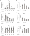
MEF knockout (KO) cells lacking each ER stress sensor or marker were used to determine the pathway through which LF regulates the ER stress. mRNA expression levels of IL6 were normalized by β-actin in the same samples.
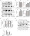
Expression level of the proinflammatory marker IL6 mRNA, were somewhat changed by LF in wt MEF; however, the variations were not dose-dependent (Fig. 4A). When stimulated with TG, significant induction of IL6 was observed, as compared to the control group (Fig. 4A). The fold change of IL6 by TG stimulation were significantly augmented in the range 12.5–50 µg/mL concentration of LF (Fig. 4B). However, in MEF cells lacking XBP1, although LF itself exerted some effect on the IL6 mRNA expression, it was not dose-dependent. Conversely, treatment with TG significantly induced IL6 mRNA expression levels at all LF concentrations (Fig. 4C). However, no significant change of IL6 induction by LF with TG stimulation was observed in the absence of XBP1, suggesting regulation through an XBP1-dependent inflammatory pathway by LF (Fig. 4D). Fig. 4E and 4F reveal that IRE1α KO MEF cells show no significant changes or notable patterns in IL6 mRNA level after exposure to either LF or TG, suggesting an IRE1α-dependent inflammatory pathway. In summary, these results show that LF increases the proinflammatory response through an IRE1α-XBP1-dependent manner in the presence of ER stress between the ranges 12.5–50 µg/mL LF.
Expression of ER stress markers in MEF KO cells
Our previous experiment reveals that LF regulates the inflammatory response via the IRE1α-XBP1-pathway on ER stress mechanism. Therefore, we next investigated the effect of LF on ER stress hallmarks BiP and CHOP in MEF KO cells.
In Fig. 5A, we present the ER stress hallmarks affected by LF. As expected, TG induces the expression of BiP, a representative ER stress marker, in all MEF cell lines (Fig. 5A). TG also induces CHOP, a representative apoptosis and ER stress marker, in all the MEF cell lines except XBP1 KO MEF. The relative intensity of the CHOP bands was calculated using the Image J software (Fig. 5B). We observed that high concentration of LF (100 µg/mL) inhibited the CHOP mRNA expression induced by TG in wt MEF and PERK KO MEF, as compared to XBP1 KO MEF and IRE1α KO MEF. This pattern is similar to expression levels of IL6 in wt MEF, and IL8 and XBP1s in polarized Caco-2 cells. This result therefore suggests that LF activates the survival mode of ER stress mechanism in an IRE1α-XBP1-dependent manner, but with no involvement of the PERK pathway (Fig. 5B).
Since LF regulates the proinflammatory response via XBP1 pathway, we further examined the expression of XBP1s by applying qRT-PCR. Fig. 5C shows that TG significantly induces the expression of XBP1s mRNA in wt MEF. In the absence of TG, the expression level of XBP1 mRNA by LF itself was not significantly changed, except at 100 µg/mL concentration of LF. However, the fold change of XBP1s mRNA induced by TG significantly decreases in a dose-dependent manner, which suggests that LF dose-dependently inhibits XBP1s in the presence of ER stress (Fig. 5D). The RT-PCR data of Fig. 5E confirms the qRT-PCR results presented in Fig 5D. The unspliced form of XBP1 (XBP1u) is seen as the upper band and the spliced form of XBP1 (XBP1s) is the lower band. Note that TG treatment shows the lower molecular weight of band (XBP1s) while the negative control shows only the higher molecular weight of band (XBP1u) in wt MEF. Since RT-PCR is not as sensitive as qRT-PCR, exposure to 100 µg/mL LF reveals only a weak band. Pretreatment of IRE1α KO MEF cells with LF showed no induction of XBP1s by TG, since IRE1α is upstream of the XBP1 molecule, and XBP1u cannot be cleaved to the spliced form in the absence of IRE1α. PERK KO MEF cells treated with LF were observed to induce XBP1s by TG, but the effect was not dose-dependent with respect to LF concentration (Fig. 5E, lower panel). Therefore, in the absence of PERK and IRE1α, no changes were observed in the levels of XBP1s after LF exposure. This suggests that LF inhibits XBP1 splicing in the presence of ER stress. Also, these results confirm the data presented in Fig. 5A, that LF does not exert its effect in regulating the inflammatory response or ER stress via the PERK pathway.
Taken together, our results indicate that 12.5–50 µg/mL of LF induces IRE1α-XBP1-dependent inflammation and inhibits XBP1s for the survival pathway; however, above 50 µg/mL, LF alters to the opposition effect by inhibiting the proinflammatory response and inducing CHOP expression, resulting in apoptosis. These are very interesting results that reveal the dose-dependent dual effect of LF.
Changes on the cell surface after treatment with LF or TG
Since the first reports of SICM imaging [37], multiple studies have reported SICM imaging of various biological samples, including cultured cells [38394041]. These previous findings indicate that SICM is a useful tool for imaging the surface topography of living and fixed cultured cells at nanoscale resolution under liquid. We utilized this new imaging technique to examine the condition of cells in experiments and the effects of any reagent (namely, LF in this study) on the surface morphology of the single cell. We obtained excellent image quality of SICM for fixed single MEF cells, thereby corroborating its potential for future studies. SICM image of intact MEF cells under various conditions indicate that the KO of XBP1 or IRE1α, and treatment of LF or TG, does not have any acute detrimental effects on the cells (Supplementary Fig. 1A). However, surprisingly, the surface of IRE1α KO MEF shows increased cytoskeleton polymerlike structures as compared to other KO MEF cell lines, indicating that knock-out IRE1α (which is ER the transmembrane sensor protein) possibly has some effect on the morphological changes, and this requires further investigation (Supplementary Fig. 1B).
DISCUSSION
LF found in China mainly has a sweet taste. Dried LF has long been used by the ancient Chinese to help the function of eyes, liver, kidneys and lungs. Furthermore, scientific studies for the Lycium barbarum fruit has proven its beneficial effects on immunomodulation, hypoglycemia, hypolipidemia, antiaging and antitumor properties [424344].
In this study, we focused on LF exerting its protective function on the gut barrier as well as against inflammation and ER stress in the intestine of humans. Our results indicate that LF protects the intestinal barrier significantly, both in the presence and absence of an inflammatory environment (Fig. 2). Although it was not statistically significant in Caco-2 cells, both MEF cell lines and Caco-2 cells showed a similar pattern of proinflammatory response along with ER stress response. The low dose of LF increased the proinflammatory response in the presence of ER stress, while at high concentrations exceeding 50 µg/mL showed no change. We believe this finding reveals a biological phenotype of the LF effect. Moreover, this effect is XBP1 dependent, implying that LF regulates inflammatory response by XBP1 through the IRE1α-XBP1 pathway followed by inhibiting ER stress and further inflammation.
As seen in Fig. 5C, the highest concentration of LF (100 µg/mL) itself induces XBP1s significantly, even in the absence of TG. We therefore conclude that higher concentrations of LF by itself are capable of inducing ER stress or general toxicity. This might be due to the survival/protection mode of the cell by activating ER stress and other protection mechanisms. However, this phenomenon needs to be followed up by further investigations.
Taken together, our results show the beneficial effect of LF on human intestine by exerting a protective function on the gut barrier, and regulation of inflammation induced by ER stress in an IRE1α-XBP1-dependent manner. In addition, AFM data shows no morphological changes by LF on MEF cells, suggesting that LF does not change or interfere with the activity with respect to cell surface morphology. However, it is interesting to note that in the absence of the ER transmembrane protein IRE1α, the cell surface is altered. Further detailed mechanistic studies are required to confirm our data regarding the influence of IRE1α on the cell surface plasma membrane, including microtubules.
 XML Download