

 PDF
PDF ePub
ePub Citation
 Print
Print


Introduction
Most chemotherapeutic agents for the treatment of cancer can destroy tumors and arrest cancer progress [1] but cancer treatment may damage healthy cells and tissues. Thus, new anticancer drugs from natural products are expected to play an important role in the development of more effective and safer agents to inhibit the onset of cancer [1]. Roland et al. [2] first isolated the anticancer factor calvaine from the basidiomycete Calvatia gigantea, which has long been used as a medicinal food. Almost 50 species in 20 generi of mushrooms have been identified as having anticancer activity [3]. The Chaga mushroom (Inonotus obliquus) has been used medicinally for treating cancer in several regions of the world [4].
Many studies have reported that I. obliquus contains excellent bioactive compounds [5,6]. 3β-Hydroxy-lanosta-8,24-dien-21-al has been identified from I. obliquus [7], and a high-molecular-weight, water-soluble, lignin-like substance from edible mushrooms of I. obliquus has been reported to inhibit the protease of type-1 human immunodeficiency virus [8]. Inotodiol isolated from the sclerotia of I. obliquus has been reported to have an inhibitory effect in a two-stage carcinogenesis test on mouse skin using 7,12-dimethyl-benz[α]anthracene [9]. Lee et al. [10] reported that a hot-water extract of I. obliquus exerts inhibitory activity against the proliferation of human colon cancer cells (HT-29). We recently identified subfractions 1 and 2 as 3β-hydroxyl-lanosta-8,24-dien-21-al and inotodial, respectively and have reported that the compounds have antimutagenic and antioxidative activities [11]. We also found that subfraction 3 has antimutagenic and antioxidative activities (unpublished results). Oxidative stress and mutagenic activity play important roles in the cancer process [12]. Thus, the possibility exists that subfractions 1, 2, and 3 also have anticancer activity in vitro against the proliferation of human cancer cells and in vivo against sarcoma growth in a mouse model. There may also be anticancer activity of subfractions containing pure compounds of I. obliquus extract in Balbc/c mice bearing Sarcoma-180 cells and in various cancer cell lines that have not been examined. Therefore, we hypothesized that the subfractions 1, 2 and 3 containing pure compounds (3β-hydroxy-lanosta-8,24-dien-21-al, inotodiol and lanosterol, respectively) separated from I. obliquus would inhibit tumor growth in mice bearing Sarcoma-180 cells (S-180) and as well as the growth of human carcinoma cell lines. The objective of this study was to test the activities of the three subfractions isolated from I. obliquus against the proliferation of cancer cells and on solid tumor inhibition as a trial for the development of novel functional anticancer medicinal food and anticancer drugs. In this study, we contributed more information about natural anticancer compounds separated from I. obliquus for the prevention or/and treatment of human cancer.
Materials and Methods
Chemicals and reagents
Fetal bovine serum (FBS), 3-[4,5-dimethylthiazole-2-yl]-2,5-diphenyltetrazolium bromide (MTT), and dimethylsulfoxide (DMSO) were purchased from Sigma Chemical Co. (St. Louis, MO, USA). RPMI1640 medium, penicillin and streptomycin (PEST), and trypsin-ethylenediaminetetraacetic acid (T-EDTA) were obtained from GibcoBRL (Gaithersburg, MD, USA). All solvents used were analytical grade.
Preparation of three subfractions from I. obliquus extracts
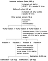
The subfractions from I. obliquus extracts were collected by the method of Ham et al. [11]. The I. obliquus specimen used in this study was imported from Russia, dried, powdered, and stored at -20℃. The powdered specimen (500 g) was extracted twice with 3 L of methanol (99.8%) at 45-50℃ for 3 h. The methanol fraction (45 g) was then rotary evaporated, emulsified, dissolved in water, and extracted three times with ethyl acetate (H2O:EtOAc, 5:7, v/v). The ethyl-acetate fraction (12 g) was obtained after rotary evaporation, and the dried extract was reconstituted in methanol. This stock solution was subjected to vacuum chromatography (N-2N; Eyela, Tokyo, Japan) with silica gel (60 g, 230-400 mesh) mixed with dichloromethane. The mobile phases were dichloromethane (A) and methanol (B), with a gradient of increasing methanol (0-100%) in dichloromethane and re-equilibration of the column with 100% A for 3 min prior to the next run (Fig. 1). The success of the fractionation was confirmed by thin-layer chromatography (TLC). Based on the Ames test and the DPPH method, amongst the three ethyl-acetate-extract fractions, VCHG-fraction 1 was the most bioactive fraction in the antimutagenic and antioxidant effects [5,6]. Thus, the VCHG-fraction 1 was selected and subjected to column chromatography using a reversed-phase column (ODS-C18) containing LiChroprep RP-18 (25-40 µm) mixed with H2O: MeOH (1:30, v/v) in a gradient program ranging from 30% to 100% methanol (mobile phase A: methanol; mobile phase B: ultra-pure water). Four fractions were isolated, of which fraction 2 exhibited the most bioactivity. Fraction 4 also had strong bioactivity. Fraction 2 and fraction 4 were further fractionated by chromatography with a normal-phase column (2.4 cm×15 cm) containing silica gel (200 g, 70-230 mesh; Merck Co., Darmstadt, Germany) mixed with dichloromethane and eluted with a mobile-phase gradient ranging from 100% dichloromethane to 50% methanol. The flow rate was 1.0 ml/min (Fig. 1). This resulted into two subfractions from fraction 2, designated as subfractions 1 and 2, both of which showed bioactivity. In our previous study, subfractions 1 and 2 were identified as 3β-hydroxy-lanosta-8,24-dien-21-al and inotodiol, respectively [11]. Subfraction 3 was obtained from fraction 4 (Fig. 1). The compound in subfraction 3 was crystallized and characterized by determining its mass spectrometry (MS) (JEOL), 1H nuclear magnetic resonance (1H NMR), and 13C nuclear magnetic resonance (13C NMR) (Bruker DRX 500 MHz) spectra.
Cancer cell and normal cell lines
The human cancer cells used in this experiment were lung carcinoma A-549 cells, breast adenocarcinoma MCF-7 cells, stomach adenocarcinoma AGS cells, Sarcoma-180 cells, and cervical adenocarcinoma HeLa cells. These, and the normal transformed primary human embryonic kidney (HEK) 293 cell line, were obtained from the Korean Cell Line Bank (Seoul, Korea). All cells were cultured in RPMI 1640 medium supplemented with 10% FBS and 1% PN/ST. The cells were incubated at 37℃ in a humid atmosphere containing 5% CO2.
Anticancer activity in vitro
The cytotoxic potential of the subfractions was evaluated by the MTT assay [12] against four human tumor cell lines: A549, MCF-7, AGS, and HeLa. The inhibitory activity against the proliferation of cancer cell lines was compared with that against the normal transformed primary human 293 cell line. The cells were seeded in 96 wells at a density of 5×10⁴ cells/ml and cultured for 24 h in RPMI 1640 medium with 10% FBS and 1% PEST at 37℃, 5% CO2. The three subfractions separated from I. obliquus were dissolved in DMSO, and the stock solutions were diluted with RPMI 1640 medium (DMSO concentration < 0.01%). The DMSO was added to all test solutions at a final concentration of 0.01%. The control was the well containing 0.01% DMSO only. Various concentrations of the subfractions (0, 62.5, 125.0, 187.5, and 250.0 µg/ml) were added to each culture well and incubated for 48 h. The supernatants were then removed by aspiration, and MTT (5 mg/ml) was added to each culture well (one-tenth of the original culture volumes). After incubation for 4 h, the medium was removed, and the cells were allowed to dry for 30 min. The precipitates were then solubilized in DMSO with shaking for 30 min. The extent of the reduction of MTT was quantified by measuring the absorbance at 570 nm.
In vivo anticancer assays
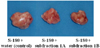
Anticancer activity in vivo against sarcoma growth in a mouse model was examined using the method of Ham et al. [13] and Maeda et al. [14] with minor modification. Briefly, Balbc/c mice (six-to seven-weeks-old) were obtained from Samtaco (Gyeonggi-do, Korea), randomly divided into the control and six experimental groups, and provided with water and normal chow. Animals were housed in an air-conditioned room at 21-26℃ with 45-55% moisture and a 12-hour dark/light cycle. The mice were treated in accordance with Kangwon National University guidelines for the care and use of laboratory animals. The Sarcoma-180 cell line was cultured for 7-10 days in the abdominal cavity of a Balbc/c mouse, and the cultured cells were harvested with the peritoneal fluid and centrifuged at 1,200 rpm for 10 min in phosphate-buffered saline (PBS). The separated sarcoma cells were floated in PBS and centrifuged, and their concentration was adjusted to 1.0×10⁶ cells/ml. The cells (0.2 ml) were implanted subcutaneously in the left groins of mice. After 24 h, the mice were fed normal chow supplemented with water (control group), 0.1 mg subfraction 1 per mouse per day (S-180+subfraction 1A), 0.2 mg subfraction 1 per mouse per day (S-180+subfraction 1B), 0.1 mg subfraction 2 per mouse per day (S-180+subfraction 2A), 0.2 mg subfraction 2 per mouse per day (S-180+subfraction 2B), 0.1 mg subfraction 3 per mouse per day (S-180+subfraction 3A) or 0.2 mg subfraction 3 per mouse per day (S-180+subfraction 3B) for 20 days. The mice were fed normal chow for 7 days after the last oral administration of subfractions and then euthanized with an ip injection of Avertin (Sigma-Aldrich Co., St. Louis, MO, USA) [15] on the 27th day after cancer-cell implantation. Solid tumors were removed and weighed, and the tumor-growth-inhibition ratio (I.R.) was calculated using the following formula:
I.R. (%) = (Cw-Tw)/Cw×100
Cw: Average tumor weight of the control.
Tw: Average tumor weight of the experimental group.
Statistical analysis
The data were expressed as means ± SD. Differences between the groups were determined by one-way Analysis of Variance using the SPSS package (Version 10.0; SPSS, Chicago, IL, USA). The level of statistical significance was set at P<0.05. The comparisons of cancer cell growth inhibition in vitro and solid tumor growth inhibition in vivo among the groups were performed using Duncan's multiple range test [15].
Results
Inhibition of cancer cell growth in vitro
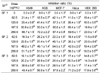
As shown in Fig. 1, the ethyl-acetate fraction from I. obliquus was separated into three fractions by vacuum chromatography. VCHG-fraction 1 was further separated into four fractions by reversed-phase (ODS-C18) column chromatography, of which the second fraction showed the greatest in vitro anticancer activity. Fraction 4 also had inhibitory activity against the proliferation of human cancer cells (data not shown). Fractions 2 and 4 from VCHG-fraction 1 were, in turn, separated by normal-phase silica-gel column chromatography into subfraction 1, subfraction 2 and subfraction 3, respectively. Each of the three subfractions yielded a single spot by TLC, confirming single-compound purity (data not shown). Purity was also confirmed by MS, 1H NMR, and 13C NMR analyses. Each of compounds 1, 2, and 3 (subfractions 1, 2, and 3) had a purity of above 99.5%. The pure compounds were assayed against human lung carcinoma A549, breast adenocarcinoma MCF-7, stomach adenocarcinoma AGS, and cervical adenocarcinoma HeLa cells and normal HEK 293 cell line.
Exposure of normal HEK 293 cell culture to 0-250 µg/ml of subfractions for 48 h resulted in low cytotoxicity (Table 1), but high cytotoxicity was observed at a concentration of 300 µg/ml subfractions (data not shown). Thus, to avoid high cytotoxicity in normal cells from subfractions, 0-250 µg/ml subfractions were chosen for in vitro study.
As shown in Table 1, subfraction 1 was more active than subfraction 2 or subfraction 3 against the selected tumor cell lines A549, AGS, MCF-7, and HeLa. Subfractions 1, 2, and 3 (250 µg/ml) inhibited the growth of A549 cells by 66.7%, 51.7%, and 48.4%, respectively, and inhibited that of AGS cells by 72.2%, 51.0%, and 56.9%, respectively (Table 1). Subfractions 1, 2, and 3 (250 µg/ml) inhibited the growth of MCF-7 cells by 67.4%, 55.1%, and 37.6%, respectively, and inhibited that of HeLa cells by 69.5%, 58.3%, and 71.2%, respectively (Table 1). In contrast, the subfractions inhibited the growth of normal HEK 293 cells by less than 20% (Table 1). The results indicate that these subfractions inhibit cancer cell growth and have low cytotoxicity against normal cells.
Solid tumor growth inhibition
In our previous study, after oral administration with 0, 0.2, 0.4, 0.8 or 1.6 mg ethyl acetate extract from I. obliquus per mouse per day, no genotoxic activity was observed, and the extract inhibited the genotoxic activity by N-methyl-N'-nitro-N-nitrosoguanidine in a dose-dependent manner [6]. Thus, to avoid toxicity from the subfractions, oral administration of 0.1 mg per mouse per day and that of 0.2 mg per mouse per day were chosen for in vivo experiment.
The average tumor weight of the Sarcoma-180-cell-bearing mice after water feeding (control) was 4.02 ± 0.19 g, whereas that of the mice after subfraction 1 administration at low level (0.1 mg/mouse per day, S-180+subfraction 1A) was 3.06 ± 0.22 g and that of the mice after subfraction 1 feeding at high level (0.2 mg/mouse per day, S-180+subfraction 1B) was 2.67 ± 0.34 g (Table 2). Thus, subfraction 1 at high level inhibited tumor weight by 33.71% of the control tumor weight, and treatment following tumor transplant resulted in a dose-dependent reduction of tumor weight in comparison with the untreated control (Fig. 2). Subfraction 2 at high level (0.2 mg/mouse per day) and subfraction 3 at both low and high levels (0.1 mg/mouse per day and 0.2 mg/mouse per day, respectively) also inhibited solid tumor growth significantly (Table 2). Subfraction 1 showed much greater inhibition of tumor growth than subfractions 2 and 3 in the mice bearing Sarcoma-180 cells, in agreement with our in vitro results.
Structural analysis of I. obliquus subfraction 3
As shown in Fig. 1, the subfractions containing pure compounds were obtained after reversed-phase (ODS-C18) column chromatography of the ethyl-acetate extract of I. obliquus, followed by normal-phase silica-gel column chromatography. In our previous study, subfractions 1 and 2 were identified by MS, 1H NMR, and 13C NMR analyses as 3β-hydroxy-lanosta-8,24-dien-21-al and inotodiol, respectively [11].
Inotodiol and lanosterol have similar chemical structures. Inotodiol has an OH group at position 22 (C-22) of lanosterol [9]. The chemical structure of the compound in subfraction 3 was determined by comparing the MS, 1H-NMR, and 13C-NMR spectra (data not shown) to published library spectra and spectra in the literature [7,9,15,16]. These analyses revealed that subfraction 3 is lanosterol. Thus, 3β-hydroxy-lanosta-8,24-dien-21-al, inotodiol, and lanosterol are bioactive compounds of I. obliquus with anticancer properties.
Discussion
This is the first study to report an antitumor effect of three subfractions (3β-hydroxy-lanosta-8,24-dien-21-al, inotodiol, and lanosterol) from I. obliquus extracts in the Sarcoma-180-cell-bearing mice and various cancer cell lines.
All of the subfractions showed significant inhibitory activity against the proliferation of the selected cancer cell lines (A549, AGS, MCF-7, and HeLa), but these subfractions have low cytotoxicity against normal cells. The subfractions inhibited cell growth in a dependent manner. Subfraction 1 was more active than subfraction 2 and subfraction 3 against the selected cancer cell lines. In our earlier study, ethanol extract from the I. obliquus showed high inhibition of the proliferation of A549, AGS, and MCF-7 cancer cells [5]. In another recent study, Lee et al. [10] reported that a hot-water extract of I. obliquus exerted inhibitory activity against the proliferation of human colon cancer cells (HT-29), in good agreement with our results.
Subfractions 1, 2 and 3 significantly inhibited tumor growth in mice bearing Sarcoma-180 cells as compared with the control mouse tumor, and subfraction 1 showed greater inhibition of tumor growth than did subfractions 2 and 3, in agreement with our in vitro results. Polysaccharides extracted from mycelia of I. obliquus not only reduce tumor size [17], but also lower elevated blood glucose [18]. Nakata et al. [9] have reported that the most abundant triterpene in I. obliquus, inotodiol, exhibits potent antitumor-promoting activity in a two-stage carcinogenesis test on mouse skin. This observation is consistent with our observations. Thus, subfractions of I. obliquus extracts have a strong anticancer effect and may be useful as an ingredient in functional anticancer food.
In this and previous studies, subfractions 1, 2 and 3 were identified by MS, 1H NMR, and 13C NMR analyses as 3β-hydroxy-lanosta-8,24-dien-21-al, inotodiol [11] and lanosterol, respectively. Our observations are consistent with those of other studies [7,9,11,15,16,19] reported that inotodiol had an in vivo antitumor effect against P388-bearing mice. Inonotus obliquus appears to have an abundance of triterpenes, including lanosterol and inotodiol [9]. A chloroform extract from I. obliquus has an antiproliferative effect on human cancer cell lines [19], and inotodiol is the main constituent of this chloroform extract [9]. Kahlos et al. [18] reported that lanosterol and 3β-hydroxylanosta-8,24-dien-21-al also inhibit proliferation of MCF-7 adenocarcinoma cells.
Lee et al. [10] showed that the hot water extract of Inonotus obliquus would be useful as an antitumor agent via the induction of the apoptosis and inhibition of the growth of cancer cells through up-regulation of the expression of proapoptotic proteins (Bax and caspase-3) and down-regulation of antiapoptotic proteins (Bcl-2). The water extract of Inonotus obliquus mushroom exhibited a potential anticancer activity against B16-F10 melanoma cells in vitro and in vivo through the inhibition of proliferation and induction of differentiation and apoptosis of cancer cells [20]. Nomura et al. [19] reported that Inotodiol, from Inonotus obliquus, inhibits cancer cell proliferation through apoptosis induction by activating caspase-3. However, the mechanisms on anticancer activity of 3β-hydroxy-lanosta-8,24-dien-21-al and lanosterol have not been examined.
In conclusion, the three subfractions (3β-hydroxy-lanosta-8,24-dien-21-al, inotodiol, and lanosterol) inhibited in vitro proliferation of various human cancer cell lines. The three subfractions also had in vivo antitumor effects on Sarcoma-180-cell-bearing mice. 3β-hydroxy-lanosta-8,24-dien-21-al, inotodiol, and lanosterol may therefore be interesting compounds for the development of novel anticancer drugs and functional anticancer food. However, further human studies are needed to determine whether dietary feeding of 3β-hydroxy-lanosta-8,24-dien-21-al, inotodiol, and lanosterol can reduce tumor growth as observed in the present in vitro and in vivo studies. We also need mechanistic studies on 3β-hydroxy-lanosta-8,24-dien-21-al and lanosterol.

 XML Download