

 PDF
PDF Citation
 Print
Print


INTRODUCTION
The term “atopic dermatitis” (AD) is already fairly common in the equine veterinary literature [1]. The diagnosis of equine AD is based on a compatible history, clinical signs (recurrent pruritus and/or urticaria) and exclusion of other differential diagnoses, such as ectoparasite infestations, insect bite hypersensitivities and possible adverse food reactions [23]. Thus far, history and intradermal testing (IDT) are gold standards in etiological diagnosis [4], but serological allergy testing (SAT) has recently been developed to detect specific IgE (sIgE), replacing the more difficult-to-perform in vivo test and have become useful tools for the identification of allergic factors [5]. The reliability of in vitro tests based on the determination of sIgE is of pivotal importance in the selection of relevant allergens for specific immunotherapy [6]. New techniques based on immunoenzymatic multiple allergen simultaneous testing (MAST) are widely used worldwide in the fields of medicine and veterinary medicine, mainly in European and Asian countries, but so far limited data are available from atopic horses. MAST can detects many kinds of specific allergen sIgE at one time with small serum volume, avoiding the cost of horse transportation to place of test performing and a procedure of multiple skin injections [7891011]. Knowledge of the extent of agreement between in vivo and in vitro test becomes particularly important when sIgE is the diagnostic test of choice due to contraindications for IDT.
Storage mites (SMs) and house dust mites (HDMs) are two of the most important allergen sources that cause severe forms of respiratory and skin allergy in horses [12]. The presence of HDMs has been confirmed in the equine environment [13]. The purpose of this study was to evaluate the reliability and diagnostic accuracy of MAST using equine monoclonal antibody mite panel compared with IDT in the etiological diagnosing of AD in horses with mite allergy by assessing the agreement between both test results.
MATERIALS AND METHODS
Criteria for animal selection
All tests were performed on fourteen atopic Malopolski horses (9 females, 5 males) with an age range of 7–16 years (median age, 11 years) from 2009–2017. Client-owned horses with suspicion of allergy admitted to the Sub-Department of Clinical Diagnostics and Veterinary Dermatology at the University of Life Sciences in Lublin (Poland, referral clinic), for evaluation of recurrent pruritus were included. All horses had clinical symptoms of AD and a history of recurrent perennial pruritus affecting the mane tail, head, thorax area, the dorsal and sometimes the ventral midline. Other causes of skin diseases were ruled out using standard diagnostic techniques (skin scraping, cytology, microbiological culture, elimination diet and appropriate therapy, deworming). Corticosteroid-responsive pruritus were observed in all horses. During the summer horses were kept on pastures with access to a shelter and during the winter in the stables in the same rural area. Prior to all scheduled tests no corticosteroids, antibiotics or antihistamines were discontinued for at least 4 weeks. Local ethical committee approved all investigations involving the use of animals.
Serological tests
The sIgE was measured from serum samples which were obtained between June and November by venipuncture of the jugular vein and sIgE measurements were performed. The blood (5 mL) was centrifuged for 10 min at 4,500 g and serum samples were kept cool at 4°C without any freeze-thaw cycles and removed 10 min before test was performed (2 to 10 h after blood collection). According to the manufacturer's instructions the allergen-specific IgE concentrations were determined in sera using 15 individual allergens, including mite whole-body extract (WBE) (Tyrophagus putrescentiae, Acarus siro, Lepidoglyphus destructor, Dermatophagoides pteronyssinus, Dermatophagoides farinae; Polycheck Allergie NF Horse Panel 20, BioCheck GmbH, Münster, Germany) and five allergen mixtures. The strips were incubated sequentially with horse serum, biotinylated monoclonal anti-equine IgE, anti-anti-equine IgE, avidin-alkaline phosphatase and substrate. The resulting processed strips were dried and scanned on a flatbed scanner. Image analysis software (BioCheck GmbH) detected the densities of each band, along with the densities of a series of control bands of graded density and expressed the results in arbitrary units/mL (U/mL). The reactions indicate an allergy classes were interpreted as negative (NR)-class 0 (< 1 kU/L), mildly positive (MPR)-class 1 (1.0–2.0 kU/L), moderately positive (MR)-class 2 (2.0–20 kU/L) and strongly positive (SR)-class 3 (> 20 kU/L) as per manufacturer's guidelines.
Intradermal tests
IDTs were performed by one investigator in autumn and winter (between September and December) in unsedated horses in the middle lateral region of the neck. After hair clipping seven injections of 0.1 mL containing mite WBE were administered intradermally using a 25-G needle—the SMs Tyrophagus putrescentiae, Acarus siro, and Lepidoglyphus destructor and HDMs Dermatophagoides pteronyssinus and Dermatophagoides farinae and other 15 (pollen, insect, molds) were included as test antigens (Agroskin RTU 20, Agrolabo Horse Panel, Scarmagio, Italy). Histamine (1:1,000 w⁄v) and phosphate buffer were used as a positive and negative control. The diameter and firmness of the wheals were evaluated semiquantitatively at 0.5 h and 4 h using a 5-gradual scale: 0 = no reaction, +⁄− = very flat reaction with a poorly defined contour, + = reaction with slight thickness, ++ = reaction with evident thickness and +++ = reaction with identical thickness as histamine [14].
Statistical analysis
The diagnostic performance of the tests was assessed using receiver operating characteristic (ROC) curve analysis. The area under curve (AUC), sensitivity (Se), specificity (Sp), positive predictive value (PPV), negative predictive value (NPV) and accuracy (Acc) of MAST were analyzed compared to IDT as the gold standard for each allergen. The optimal cut-off points were determined from the ROC curves, with an equal weight of costs of wrong decisions by the Youden index. Spearman rank correlation test was used in the analysis of associations between IDT and MAST. Cohen's kappa coefficient was used to evaluate multiple levels of agreement between IDT and MAST for dichotomic parameters (DPs) (qualitative, positive-negative) and semiquantitative parameters (grade of the intensity of the test reaction [GIR]): less than 0, disagreement; 0–0.2, slight agreement; 0.2–0.4, fair agreement; 0.4–0.6, moderate agreement; 0.6–0.8, substantial agreement; and 0.8–1, almost perfect agreement. Data analyses were performed using STATISCTICA version 13.3 (TIBCO SOFTWARE Inc., Ireland).
RESULTS
Using a mean IDT cut-off diameter of at least + and an IgE cut-off value ≥ 1.0 kU/L for MAST, the most prevalent positive reactions and highest mean values in all performed tests were reported against T. putrescentiae (IDT, 79%; MAST, 79%) and D. farinae (IDT, 71%; MAST, 79%). The lowest intensity positive response was observed against A. siro (IDT, 43%) and L. destructor (MAST, 50%) (Table 1).
Table 1
Analysis of IDT results (+, ++, and +++) and IgE concentrations in sera (kU/L) showing the number, percentage of positive reactions, mean values and ranges on the MAST in horses with AD

ROC analysis showed that sIgE to A. siro had the best diagnostic performance (AUC = 0.969), followed by D. pteronyssinus (AUC = 0.844), D. farinae (AUC = 0.813), T. putrescentiae (AUC = 0.803) and L. destructor (AUC = 0.467) (Fig. 1). Using the statistical optimal cut-off (based on Youden's index) for sIgE, which was determined from the ROC analysis, a clinical sensitivity and specificity of 86% and 100% (A. siro), 73% and 100% (T. putrescentiae), 44% and 60% (L. destructor) were obtained for SMs and a clinical sensitivity and specificity of 80% and 75% (D. farinae) and 89% and 80% (D. pteronyssinus) were showed for house mites. The diagnostic accuracy of MAST varied between 50% for L. destructor and 90% for A. siro (Table 2).
Fig. 1
ROC curves for intradermal test and allergen-specific IgE to mites based on multiple allergen simultaneous test analysis of sera in horses with atopic dermatitis.
ROC, receiver operating characteristic.
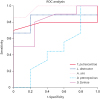
Table 2
Diagnostic performance of specific IgE measurement in response to mite allergens calculated from MAST compared to IDT data

Manufacturer and optimal cut-off points were determined using the Youden index.
Man, manufacturer; Opt, optimal; Se, sensitivity; Sp, specificity; PPV, positive predictive value; NPV, negative predictive value; Acc, accuracy; AUC, area under the curve.
Significant values are indicated by *p < 0.05, **p < 0.01, ***p < 0.001.
A significant positive correlation between IDT and MAST was found for A. siro (rS = 0.870; p = 0.00005) and D. farinae (rS = 0.657; p = 0.011). Agreement between IDT and MAST was globally weaker when we graded the intensity of both test reactions than when we used DPs (positive-negative). Significant agreement was obtained for 2 of 5 allergens: A. siro (κ = 0.569) and D. fariane (κ = 0.485) for GIR and for 3 of 5 allergens: D. pteronyssinus (κ = 0.689), A. siro (κ = 0.571), D. fariane (κ = 0.432) for DP (Table 3).
Table 3
Correlation and agreement analysis between sIgE concentrations (kU/L) and IDT results

DISCUSSION
In this study, we evaluated the performance of a new immune-enzymatic technique for allergen-specific IgE determination in horses with AD. To our knowledge, this is the first study comparing MAST with IDT in horses with storage and domestic mite hypersensitivity. In human medicine, such tests (MAST) have been studied using approved in vivo skin prick tests (SPTs) and in vitro (Phadia, sIgE) methods [15]. Despite current intensive research in the field of immunology, which is promising for further progress in recombinant allergen development [16], most standardized serological tests are based on WBE allergens. Mite recombinant allergens, with the exception of D. pteronyssinus (rDer p 23) and Glycophagus domesticus (rGly d 2), have not yet been presented in serological tests in horses [17]. The use of such recombinants would result in an increase in the specificity of the serological tests and reduce the incidence of false-positive reactions in horses.
In our studies, IDT and MAST showed a high percentage of positive reactions to SMs (mainly T. putrescentiae) and dust mites (D. farinae) what confirms the importance of both allergens in the development of AD in horses. Similar results were observed by Roberts et al. [18], who reported that most positive results in serological tests were obtained for D. farinae (24%) and T. putrescentiae (18%). The highest agreement for the intensity of the reaction (semiquantitatively) in certain individuals in both tests was for A. siro and D. farinae, but concordance was not demonstrated statistically for the other mites. However, the intensity of the positive reaction does not necessarily indicate a higher significance of the allergen in the etiopathogenesis of the disease. In contrast, comparing dichotomous response categories (positive and negative reactions) showed the highest level of test agreement and indicated that MAST is useful as an alternative to in vivo tests in the diagnosis of causative allergens in horses. In allergology practice, allergen selection for specific immunotherapy is determined by assessment of positive test results, geographical location and seasonality of symptoms, not always based on the strongest reactions in the test [19].
Our findings demonstrate that sIgE to A. siro, D. pteronyssinus, D. farinae and T. putrescentiae based on MAST results is good predictor of mite allergy in horses (AUC ranges 0.803–0.969) but to L. destructor showed lack of diagnostic value (0.467). Comparisons of serological test results (sIgE in sera) and IDT were previously performed in horses, mainly in the course of recurrent airway obstruction, [1819], skin hypersensitivity [2021] and insect hypersensitivity [324]. Morgan et al. [23] assessed horses with seasonal and perennial symptoms of skin hypersensitivity and urticaria, demonstrating low concordance between IgE and IDT, except for grass allergens. Lorch et al. [22] studied horses with recurrent urticaria, AD, and RAO and reported poor agreement for both tests with mite allergens. The highest agreement was shown in the ELISA test containing monoclonal antibodies in a group of horses with AD. Tahon et al. [20] did not demonstrate the relevance of the mites for the development of RAO in horses, although 50% of the horses had positive reactions with a commercial ELISA test using the FceRIa against T. putrescentiae, with values of 23% for D. farinae and D. pteronyssinus and 15% for A. siro. Positive reactions were also observed in healthy control animals. Tilley et al. [21] showed the highest compatibility between serological tests and the prick test for mites (D. farinae) among all groups of allergens. Thus, the sensitivity, specificity and agreement of the different IgE tests varied according to the group of allergens, the type of allergic disease and even individual allergen belonging to the same antigenic group what our study supports. Despite the different test results obtained in comparative studies, mites are a group of major allergens that have the highest percentage of positive responses in in vivo and in vitro tests. Our studies also support the importance of mite allergens in a group of horses with AD when the main symptom is recurrent pruritus without urticarial, food factors were ruled out as the cause of the observed clinical signs, and the horses responded well to anti-inflammatory doses of glucocorticoids. The probability of sensitivity to mites and positive reactions in such a cohort of animals is very high.
After comparing the values suggested by the manufacturer for MAST and the optimum cut-off values obtained based on the ROC curve and the Youden index, the values overlapped in two cases, but for three allergens, the optimal values of the cut-off points were higher. Similar observations concerning cut-off points have been made in evaluations of diagnostic techniques in medical [2526] and veterinary studies of recombinant allergens [18], and the ideal equine IDT cut-off threshold values for house and storage allergen dilution concentrations for an immediate type I reaction have been reported [18]. The application of ROC analysis helps in determining a suitable cut-off for each test, with the aim of approaching 100% certainty of diagnosis [26]. Our results suggest that increasing the cut-off points could improve the accuracy and specificity of MAST.
In our studies, blood was collected during the period of the highest concentration of mite allergens in the environment. These periods correlate with the occurrence of higher mite density in the horse's environment [18]. Scheduling testing during the symptomatic period is crucial for serological tests and reliability of etiological diagnosis for AD in which we can expect the presence of sIgE in sera. This allows for greater understanding of interdependence between clinical symptoms and results of serological test. However, false-negative results can occur in asymptomatic period. IDT was performed in fall and winter during asymptomatic period of the disease [27]. Though, the greatest severity of symptoms associated with allergies to dust mites occurs in summer and autumn, however, symptoms may occur year-round [18].
A critical limitation of this study was the use of IDT as empirically reference method for evaluation of newly developed analyzers. However, IDT is the official reference procedure for allergology practice with good reliability and reproducibility [1422].
In conclusion, our study confirms the usefulness of MAST using sIgE for storage and domestic mites in horses with AD. The pivotal dust mite allergens in horses are T. putrescentiae and D. farinae, which should be taken into consideration for the development of recombinants that could be used in serological tests and specific immunotherapy. High agreement between MAST and IDT applies to positive or negative reactions in atopic horses, and lower concordance occurs in GIR tests. To improve the accuracy of the test, the cut-off point for positive reactions should be increased.
 XML Download