

 PDF
PDF ePub
ePub Citation
 Print
Print


Introduction
Atopic dermatitis (AD) is an inflammatory and pruritic allergic skin disease with characteristic clinical features [11]. Aside from genetic predisposition, a number of environmental triggers cause AD in dogs and humans, and various investigations have been conducted to control hypersensitivity responses. Identification of the causative agents allows for allergen-specific immunotherapy and may also aid in potential avoiding the offending allergens [15,19,20]. Canine and human AD have been the focus of a great deal of research around the world. In humans, house dust mites (HDM), cockroaches, pets such as cats and dogs, pollen, and mold have been identified as important causative allergens [10,12,18]. Previous studies on risk factors for canine AD showed that house dust and HDM are common allergens in dogs [13,20].
Currently, the most common diagnostic methods used for identifying allergens in atopic dogs are the intradermal test (IDT) and in vitro antigen-specific IgE testing. IDT more closely reflects the natural disease pathogenesis in dogs rather than serologic IgE testing [3,19]. Allergens involved in intradermal reactivity vary according to population numbers, geography, and life styles [5]. Therefore, identifying common causative agents of allergic diseases could be helpful for understanding the temporal changes of environmental conditions and geographic differences among countries. It could present a new sight of relation between disease and environmental conditions.
Many studies of canine AD allergen distribution have been conducted over the past 20 years, but within the past five years studies on the topic have been rare. In addition, comparative research concerning common canine AD allergens in Asia has been lacking. The purpose of this study was to identify the most prevalent allergens affecting atopic dogs in Korea by IDT. We compared the results of our investigation with those from studies conducted in other countries.
Materials and Methods
Animals
Fifty-eight privately-owned dogs with spontaneous AD were enrolled in our study conducted at the Veterinary Medical Teaching Hospital of Konkuk University (Korea) from 2004 and 2008. A clinical diagnosis of AD was determined based on a combination of medical history and clinical signs according to diagnostic criteria [19]. Potential diagnoses of other skin diseases (e.g., adverse food reactions, ectoparasites, or endocrine disease) were ruled out. Routine skin examinations included skin scrapings and cytologic preparations. Secondary infections such as those caused by bacteria, Malassezia, and fungi were treated with appropriate medications prior to IDT.
Physical characteristics and clinical history
The breeds included in this study were Maltese (n = 11), Shih-tzu (n = 11), Yorkshire terrier (n = 8), Cocker spaniel (n = 6), Pekingese (n = 5), mixed (n = 5), Pug (n = 3), Pomeranian (n = 1), King Charles spaniel (n = 1), Schunauzer (n = 1), French bulldog (n = 1), Poodle (n = 1), miniature Pincher (n = 1), Chihuahua (n = 1), Fox terrier (n = 1) and Sabsari (a native Korean breed, n = 1). Twenty-six (44.8%) out of the 58 dogs were female, and thirty were male (51.7%). There was no gender predilection of AD in this study (p = 0.0690). Forty-nine of the 58 dogs (84.5%) were neutered or spayed. Ages ranged from 1 to 13 years with a mean age of 4.8 years, and more than 70% of the dogs had clinical signs of AD prior to being 3-years-old.
Allergens
Allergen extracts for IDT were purchased from Greer Laboratories (Lenoir, USA). The 39 grouped allergens used for this study were prepared according to the manufacturers' instructions. The allergen groups consisted of pollens, weeds, trees and shrubs, flowers, molds, smut, house dust, epidermal and inhalants, HDM, and insects. Epidermal and inhalant allergens included cat epithelia, cotton seeds, kapok seeds, prethrum, silk, and mixed feathers. The HDM allergens included Dermatophagoides (D.) farina and D. pteronyssinus. Insect allergens consisted of mosquitoes, two cockroach mixes, and fleas (Ctenocephaides spp.).
The prepared concentrations of the various allergens were 250 protein nitrogen units (PNU)/mL for the Rhizopus mix of molds, 100 PNU/mL for house dust, 1 : 5,000 (w/v) for the HDM allergens, 1 : 1,000 (w/v) for fleas, and 1,000 PNU/mL for the remaining allergens. All allergens were diluted with a sterile diluent (0.9% sodium chloride and 0.4% phenol) before use. The diluent was used as a negative control, and histamine phosphate (0.0275 mg/mL) was used as a positive control.
IDT
IDT was performed according to standard methods [15,19,20]. Briefly, glucocorticoid and anti-histamine therapies were discontinued for a minimum of 8 weeks and 4 weeks, respectively, prior to the IDT. The dogs were sedated with an intravenous injection of 10 µg/kg of medetomidine hydrochloride (Domitor, Finland) and the hair coat on the lateral thorax was carefully clipped to avoid subsequent skin irritation. Approximately 0.05 mL of each allergen was injected intradermally using an insulin syringe (BD Ultra-Fine, USA). The diameter of the resulting wheal was measured with a ruler 15 min after injection. Based on previous reports [15,20], a positive reaction was defined as a diameter equal to or greater than half of the diameter of the positive and negative controls. The results were subjectively graded on a scale of 0~4 according to the diameter, height, firmness, and erythema of the wheal. The negative control was graded as 0 and the positive control was graded as 4. All scores of 3 and 4 were considered to be positive results and clinically relevant.
Results
IDT
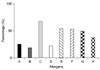
The percentages of positive reactions to allergens in each group are shown in Fig. 1 and Table 1. Only 10.3% of dogs with AD reacted solely to one allergen while the remaining animals reacted to two or more allergens. The highest rate of positive allergen reactions was against molds (67.3%). Twenty-seven dogs (49.1%) that were allergic to molds also had positive reactions to Rhizopus, the second most common reactive allergen, followed by house dust (54.5%). The epidermal and inhalant allergen group was also associated with relatively high frequencies of positive reactions (52.7%). In this group, silk (32.7%) and cat epithelia (14.5%) individually elicited a high rate of positive reaction. HDM also caused a relatively high percentage of reactions (49.1%). Reactions against individual HDM allergens (D. farina and D. pteronyssinus) were similar in prevalence (40% and 43.6%, respectively).
Twenty-eight (48.3%) dogs had positive reactions to allergens in the insect group (fleas, mosquitoes, and two cockroach mixes). Fourteen (25.5%) out of the 58 dogs had a positive reaction to flea antigens, and mosquito allergens also elicited a positive reaction in 25.5% of this group. Few positive reactions to outdoor allergens including pollen, weeds, smut, and trees and shrubs were observed.
Discussion
In the present study, two breeds of small dogs, Maltese and Shih-tzu, were most commonly afflicted with canine AD. These results may be related to the preference for small breeds by dog owners in Korea.
The IDT results of this study revealed that house dust was the allergen that most commonly produced a positive reaction (54.5%). House dust is also an important environmental allergen in humans and other animals [4,7], and is the most common allergen associated with canine AD [14,15]. However, there is no standardization for the production of house dust extract components among companies that manufacture allergens for laboratory purposes. Likewise, there is also a lack of standardization for the protocols of experiments conducted the same laboratory. This is because the components of house dust can significantly change due to the season and environmental contamination [17].
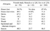
Comparisons of the frequency of reactions against causative canine AD allergens from prior studies [8,20] are shown in this study. The frequency reaction against tree and grass allergens in the present Korean study was lower than those observed in the other countries. In addition, the allergen components which were tested were similar except for a greater prevalence of mold as an indoor allergen in the present study.
Reaction to Rhizopus was relatively common in this study (n = 27, 49.1%), similar to previous results from other countries. Other studies have reported that HDM as the most common allergen of canine AD [3,16,19,20]. In addition, HDM are the main causative allergen for human atopic patients in many countries including Korea [6,9]. The present prevalence of positive IDT reactivity to HDM was relatively low compared to those found in other countries. These observations could be related to the possibility that the test antigens manufactured by different companies could contain different concentrations or possess different biological potencies [5]. Additionally, housing conditions have changed over time in Korea. Further studies are needed to investigate the factors which may affect the frequencies of reactions to causative canine AD allergens.
Outdoor allergens such as trees, weeds, and grasses were found to cause few positive reactions in dogs living in Korea. This may be related to the preference for small breeds by pet owners in Korea, where dogs are commonly housed indoors rather than outdoors, and with limited outdoor exposure of the animals. In humans, allergic reactions to indoor allergens such as HDM and cats were also significantly higher in Seoul than in a study conducted in Ankara, Turkey [16]. Improvement of indoor environments may be very important for managing canine AD in dogs residing in Seoul.
Among environmental factors, geographic and meteorological conditions contribute to the development of AD [16]. In Japan, HDM and Japanese cedar pollen are the most common allergens. Japanese cedar pollen is a very unique allergen to a particular region in dogs and humans [8]. In Korea, no allergens have yet been reported as being unique to any certain region. However, further studies may uncover specific regional differences.
Interestingly, positive reactions to flea antigens were relatively high (25.5%) in the present study. A report from the United States showed similar frequencies in reactions to insect causative allergens [20]. This may be due to the fact that northern California has a large endemic flea population [20]. On the other hand, fleas have not been an important allergen in Korea; there have been no reports of flea-related AD in dogs until recently. Korea, however, has a humid, continental climate marked by sharp seasonal changes and fleas may be endemic. Therefore, monitoring flea allergies associated with canine AD in Korea is warranted.
It is reasonable to assume that the climate of Seoul contributes to the recent increase in mosquito populations. In addition, cockroaches have been recently found to commonly cause positive allergic reactions among humans in Korea [6,13,16]. This recently discovered tendency may be related to climate changes and environmental pollution in Korea [2].
In the present study, all positive reactions on the IDT were thought to indicate causative allergens of AD in dogs. According to some previous studies, IDT cannot be used as a sole method for distinguishing atopic from normal dogs due to the appearance of clinically irrelevant reactions in normal dogs [1,7]. Such reactions could indicate a subclinical hypersensitivity state, or the necessity of factors other than mast cell-bound antibodies for the development of AD (such as defects in barrier function, abnormal mast cell function, IgE receptor mutations, or up-regulated cytokine pathways) [5]. The dogs in the present study, however, had been diagnosed with AD based on a combination of medical history and clinical signs evaluated with diagnostic criteria. Therefore, the IDT reactions of the dogs in the present study could be related to the clinical signs of AD.
In conclusion, our investigation found that the most common causative allergens for canine AD in Seoul are molds, house dust, HDM, insects, and inhalants. These results might reflect climate changes in Korea. In addition, the indoor lifestyle of the small breeds of dogs most commonly kept as companion animals may have affected the differences of reactions against various allergens compared to those observed in atopic dogs from other countries.
 XML Download