

 PDF
PDF ePub
ePub Citation
 Print
Print


Introduction
Promoting human health by enhancing natural immune protection via bioactive compounds derived from plants has recently attracted considerable scientific interest and attention [19]. There are several advantages offered by bioactive compounds extracted from plants: low toxicity; high yield and easier obtainment; and favorable physiological functions, e.g., they can improve immunomodulation and have anti-oxidative, anti-microbial, and anti-hypertensive functions [20]. Plant extracts have been widely used as biological additives and for drug development. Notably, preclinical development of bioactive natural products and their analogs as chemotherapeutic agents is required for their future clinic applications.
Betulinic acid (BA) is a pentacyclic triterpene which is found in the stem bark of the plant white birch, and also in various other plants widespread in tropical regions such as Tryphyllum peltaum, Ancistrocladus heyneaus, Diospyoros leucomelas, Tetracera boliviana, and Syzygium formosanum [7,15,25]. BA and its derivatives have been the subject of intense study with focuses on their anti-cancer effects [22,23], anti-HIV [13], anti-bacterial, anti-inflammatory [24], antimalarial [6], anti-helminthic [10], and other pharmaceutical properties [2,12]. These effects may be due to their ability to modulate immune function rather than having a direct effect on infections and on cancer cells. In addition, various bioactive materials derived from plants exhibit an immunomodulatory ability [18]. Therefore, we propose that BA may be another valuable immunomodulator. The purpose of the present study was to determine whether BA affects mouse innate and adaptive immunity, which may lay fundamental groundwork for BA-based drug development.
Materials and Methods
Chemicals and antibodies
Concanavalin A (Con A), lipopolysaccharide (LPS), trypan blue, dimethylsulfoxide (DMSO), 3-(4,5-dimethylththiazoyl-2-yl)2,5-diphenyltetrazolium bromide (MTT), and Penicillin-Streptomycin were obtained from Sigma-Aldrich (USA). RPMI-1640 was obtained from Gibco (USA), and fetal bovine serum (FBS) was purchased from Hyclone (USA). Antibodies including rat anti-mouse CD4: fluorescein isothiocyanate (FITC) / CD8: R-phycoerythrin (RPE) (DC 034), rat anti-mouse CD19: FITC / CD3: RPE (DC 035) were obtained from BD (USA). The ELISA kits for assaying IL-2, IL-6, IL-10, and TNF-α were purchased from R&D System (USA).
Preparation of BA
Plant material
The white birch bark samples were collected during spring, 2009 from Wroclaw, Poland. All the collected barks were immediately dried at 60℃ and stored in a dry and dark place.
Extraction, synthesis, and identification of BA
Fifteen g of dried bark were refluxed with 200 mL methanol for 3 h at 70℃. The methanol extract was dried under negative gauge pressure and dissolved in dichloromethane. After adding 2 M sodium hydroxide and mixing, the lower layer liquid was collected and then filtered under negative gauge pressure. The remaining substance was dissolved in ether, and water was added and mixed well. After that, the upper layer liquid was collected, filtered and fractionated with hexane and ethyl acetate (6 : 1). Betulin was obtained by silica gel column chromatography. The compound was subjected to Jones reagent oxidation to obtain betulonic acid. Reduction of betulonic acid by sodium borohydride in tetrahydrofuran provided a mixture of 3α- and 3β-hydroxyl products (5 : 95). Crystallization of the product mixture from methanol resulted in the 3β-hydroxyl product at a 75% yield.
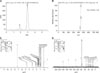
Synthetic compound was a white powder and its molecular weight was 456 by mass spectrometry (MS) (Agilent 1100 Series LC/MSD, USA). 1H-NMR spectral (Varian INOVA-300, USA) data of the compound (CDCl3, 300 MHz) is as following; δ: 0.754, 0.824, 0.934, 0.967, 0.977, 1.691 (all s, each 3H, H-23, H-24, H-25, H-26, H-27, H-30), 2.252 (m, 1H, H-19), 3.20 (dd, 1H, H-3), 4.616 and 4.744 (br s, each 1H, H-29). 13C-NMR spectral data of the compound (CDCl3, 300 MHz) is as following; δ: 38.369 (C-1), 27.377 (C-2), 78.991 (C-3), 38.842 (C-4), 55.312 (C-5), 18.26 (C-6), 34.288 (C-7), 40.658 (C-8), 50.483 (C-9), 37.187 (C-10), 20.824 (C-11), 25.469 (C-12), 38.682 (C-13), 42.412 (C-14), 29.68 (C-15), 32.129 (C-16), 56.289 (C-17), 46.875 (C-18), 49.24 (C-19), 150.386 (C-20), 30.527 (C-21), 37.011 (C-22), 27.972 (C-23), 15.324 (C-24), 16.01 (C-25), 16.109 (C-26), 14.675 (C-27), 180.526 (C-28), 109.688 (C-29), 19.351 (C-30). The identity of BA was confirmed by comparing the results of MS, 1H-NMR and 13C-NMR analysis with an authentic sample (Fig. 1).
Animals and experiment designs
A total of 112 female Kunming mice weighing 18~22 g (six weeks of age) were used in the present study. All animals were obtained from the Animal Service of Health Science Center in China. Mice were maintained on a laboratory standard diet and water ad libitum, and kept in an environment with constant temperature (23 ± 1℃) and humidity (60 ± 10%) under a 12-h light/dark cycle. All animal care procedures were carried out pursuant to the guidelines of Laboratory Animal Care (NIH Publication No. 85-23, revised in 1985; USA).
Animals were randomly classified to four groups: the control group, and 0.25, 0.5, and 1 mg/kg body weight BA-treated groups. Each experimental group consisted of seven mice. BA was administered to mice per os with 1% starch jelly for 2 weeks, and the control mice were given an equivalent amount of pure starch jelly; the total volume of the drug was 0.2 mL/20 g/mouse. Animal body weights were obtained weekly to determine the effects of BA on body weight and to adjust the administered amount of BA. After 2 weeks, the mice were anaesthetized with halothane and then sacrificed by cervical dislocation. Blood samples were collected through retro-orbital bleeding at specified time points under halothane anesthesia and assayed for cell counts, cytokines, and antibody titres.
Effects of BA on the immune apparatus index
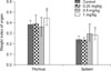
The weights of spleen and thymus were measured after being excised from the animals. The thymus and spleen indices are expressed according to the following formula: weight of organ (mg)/body weight of mouse (mg) × 100.
Lymphocyte proliferation assay
Lymphocyte proliferation was determined by MTT assay. The spleens were removed using a sterile technique and placed in sterile plates containing Hank's balanced salt solution (HBSS). The splenocytes were dissociated from the connective tissue capsule by gently pressing the organ through a 200 mesh sterile metal sieve. The sieve was rinsed with HBSS, and the suspension collected in sterile 15 mL conical tubes. Red blood cells in the cell suspension were dissolved in haemolysed solution (7 g/L NH4Cl and 2.6 g/L Tris-HCl). The resulting single cell suspension was washed twice with HBSS and centrifuged at 180 × g for 10 min. The supernatant was discarded, and the cell pellets were re-suspended in 1 mL RPMI-1640 complete medium (supplemented with 10% (v/v) FBS, 100,000 units/L of penicillin and 0.1 mg/mL of streptomycin) at a density of 5 × 10⁶ cells/mL. The purity of isolated cells was approximately 95%, and the viability of splenocytes was always more than 95% as determined by a trypan blue dye exclusion assay.
200 µL of lymphocytes were stimulated with 5 µg/mL Con A and/or 10 µg/mL LPS, and cultured in 96-well plates (Corning, USA) for 68 h at 37℃ in a humidified atmosphere of 5% CO2. Next, 15 µL of MTT (5 mg/mL) was added to each well. After a 4 h incubation at 37℃ and 5% CO2, the medium was removed and 150 µL of DMSO were added to each well. The absorbance (A) was measured on Microwell Reader (Tecan Sunrise, Austria) at 490 nm. The experiments were performed in triplicate and the results were described as an average of A.
Determination of plaque forming cells (PFC)
Sheep red blood cells (SRBC) were collected from a healthy sheep and stored in sterile Alsever's solution (components: 2.05% glucose, 0.42% sodium chloride, 0.8% tri-sodium citrate and 0.055% citric acid in distilled water) and then washed three times with 0.1 mol/L phosphate-buffered saline (PBS, pH 7.2) solution. Mice were immunized with 0.2 mL of 20% (v/v) SRBC solution (4 × 10⁸ cells/mouse) by intraperitoneal, and the number of PFC was determined on days 4 after SRBC injection. 100 µL of a splenocyte suspension (1 × 10⁶ cells/mL) and 100 µL of 10% (v/v) SRBC were mixed with 0.5 mL of 0.5% agarose and then transferred to a slide covered with a 0.5% agarose layer. After incubation at 37℃ in 5% CO2 and adequate reaction with guinea pig complement, the number of plaques was counted and expressed as those per 1 × 10⁶ viable splenocytes.
Determination of anti-SRBC antibodies in mouse serum
Anti-SRBC haemagglutinin titre was determined on day 4 after SRBC immunization. Blood was taken from the retro-ocular arteria of halothane anaesthetized mice. The sera were obtained by blood centrifugation at 3,000 rpm for 10min and inactived at 56℃ for 30 min. The total and 2-mercaptoethanol-resistant serum agglutination titres were defined by an active haemagglutination test according to the procedure previously described by Adler [1]. The titre of 2-mercaptoethanol-resistant antibody was roughly equivalent to that due to IgG in the serum, and thus the greater titre obtained without 2-mercaptoethanol was due to IgM. We found that serum from non-immunized mice didn't contain anti-SRBC antibodies.
Assay of thymocyte and splenocyte subpopulations
The mice were anaesthetized with halothane. The thymus and spleen were removed and placed in disposable Petri dishes containing sterile, ice-cold PBS. The suspended cells were released from the lymphatic organs by passing them through a nylon mesh and centrifuging them onto a layer of Ficoll 400 (Pharmacia, Sweden) / Uropolinum 75% (diatrizoate sodium and meglumine diatrizoate; Polpharma, Poland) at a 1 : 3 ratio and a density of 1.071. After centrifugation (3,500 rpm for 20 min) at 4℃, the cells were collected from the interphase and washed twice with PBS supplemented with 1% bovine serum albumin (BSA) at 4℃. After the second wash, the cells were suspended in PBS with 1% BSA at 1 × 10⁷ cells/mL. The purity of isolated cells was approximately 95%, and the viability of each cell suspension was 90~98% according to a trypan blue dye exclusion assay. The cells were re-suspended in 100 µL of a PBS solution containing 1% BSA. The thymocytes and splenocytes were stained with rat anti-mouse CD4: FITC / CD8: RPE at the dilution recommended by the manufacturer (BD, USA). The splenocytes were also stained with rat anti-mouse CD19: FITC / CD3: RPE at the dilution recommended by the manufacturer (BD, USA). The cells were incubated at 4℃ for 30 min, and washed three times with ice-cold PBS buffer. The fluorescence was analyzed using a flow cytometer (FACS Calibur; Becton Dickinson, Germany). The distribution of the thymocyte and splenocyte markers was analyzed using software (CellQuest Pro; BD, USA).
Peritoneal macrophage neutral red dye uptake assay
Peritoneal macrophage culture
Peritoneal cells were harvested 3 d after intraperitoneal injection of 0.5 mL of a starch solution (5% concentration) and isolated using 10 mL PBS with antibiotics (penicillin 10 U/mL and streptomycin 1 mg/mL). The cells were washed and plated in 100 µL RPMI-1640 medium supplemented with 10% FBS, penicillin (100 U/mL) and streptomycin (100 µg/mL) at a density of 2 × 10⁶ cells/mL in 96-well flat bottom plates. The cells were incubated for 3 h at 37℃ in an atmosphere of 5% CO2 and washed with PBS to remove non-adherent cells.
Neutral red uptake assay
After 24 h incubation at 37℃ in 5% CO2, the medium was removed, and 100 µL of 0.1% neutral red in PBS was added to each well and incubated for 3 h. The cells were washed with PBS three times, and 100 µL of a 1% acetic acid solution (v/v) in 50% ethanol (v/v) was added to each well to extract the dye phagocytised by macrophages. After rapid agitation on a microtitre plate shaker, the absorbance was read at 540 nm. Tests were performed in triplicates from three experiments.
Cytokine levels by ELISA assays
Measurement of IL-2, IL-6, IL-10 concentrations in mouse serum
Blood was collected and centrifuged at 4℃ at day 14 from mice without or with BA treatment; the serum was stored at -80℃ before assay. The IL-2, IL-6, IL-10 contents in serum were determined by ELISA according to the manufacturer's protocol.
Measurement of TNF-α concentration in macrophage culture supernatants
After purifying the macrophages, incubation was continued for 18 h and the medium was replaced with fresh medium without FBS, but containing LPS from Escherichia coli at a concentration of 2.5 µg/mL. Each culture was tested in triplicate. After 24 h of incubation, supernatants were removed and stored at -80℃. A commercial ELISA kit was used to determine mouse TNF-α in macrophage culture supernatants according to the manufacturer's instructions.
Statistical analysis
The one-way analysis of variance test and multiple comparison of Dunnett's t-test were used to evaluate the difference of parametric samples between the control and BA-treated groups, and data were expressed as the mean ± SD of the values. A p value <0.05 was considered significant difference, and p < 0.01 was considered to be an extremely significant difference between BA-treated mice and the control.
Results
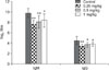
Effects of BA on the immune apparatus index
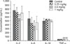
Daily oral administration of BA did not result in any mortality. No obvious signs of toxicity were observed during the time of administration. Fig. 2 shows that the thymus indices of the mice in the high dose group (1 mg/kg body weight) and the spleen indices of the mice in the medium and high dose groups (0.5 mg/kg and 1 mg/kg body weight) were increased compared to those of the control group.
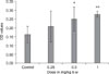
Effect of BA on proliferative response of splenocytes to Con A and LPS
We compared the general effect of BA on splenocytes proliferation. Fig. 3 shows that BA caused a significant increase in proliferative response in Con A- and/or LPS-stimulated splenocytes; in particular, proliferation of splenocytes was dramatically increased in a dose-dependent manner after stimulation with Con A and LPS together. BA-treated splenocytes cultured in absence of mitogens did not show any significant proliferative effect.
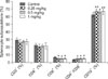
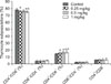
Effect of BA on the humoral response of SRBC-immunized mice
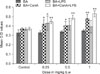
We next probed the effect of BA on the antibody response of humoral immunity in mice. From the plaque-forming cells assay, the number of PFC per 1 × 10⁶ splenocytes was enhanced by 15.9%, 24.8%, and 54.3% in BA-treated groups (0.25 mg/kg, 0.5 mg/kg and 1 mg/kg, respectively), compared with those in the control (Fig. 4), and the 1 mg/kg dose of BA had the most significant effect on PFC. Moreover, the production of total and 2-mercaptoethanol-resistant anti-SRBC serum haemagglutinins (IgM and IgG) was significantly decreased in BA-treated groups (Fig. 5). IgM was reduced by 21.8%, 17.4% and 14.5% and IgG was reduced by 24.9%, 18.8% and 15.5% in BA-treated groups (0.25 mg/kg, 0.5 mg/kg and 1 mg/kg, respectively).
Effect of BA-treated on macrophage phagocytosis
To evaluate the activity of macrophage phagocytosis, we assessed the function of phagocytic capacity of peritoneal cavity phagocytes in mice. Fig. 6 shows that the macrophage phagocytic activity was significantly increased by BA at the medium and high doses (0.5 mg /kg and 1 mg/kg) compared to the control group. No significant effects of BA on macrophage phagocytic activity were observed in mice at the dose of 0.25 mg/kg body weight.
Effect of BA on thymocyte and splenocyte subpopulations
Since BA enhanced the activities of lymphocytes possibly by altering the quantities of T and B cells or their subpopulations, we explored the effect of BA on thymocyte and splenocyte subpopulations. As shown in Fig. 7, the percentage of CD19+ splenocytes and the splenic ratios of CD4+/CD8+ were significantly increased in BA-treated mice, whereas percentage of CD8+ splenocytes was significantly decreased compared to the control group. There were no obvious changes in the percentage of total T cells (CD3+) and CD4+ T cells. As shown in Fig. 8, the percentage of CD4+CD8- thymocytes in BA-treated groups (0.25 mg/kg, 0.5 mg/kg and 1 mg/kg) was significantly increased by 8.81%, 11.37%, and 14.8%, respectively, while there were no significant changes in the percentage of CD8+CD4- and CD8-CD4- T cells, nor in the ratio of CD4+/CD8+ T cells.
Effect of BA on cytokine levels
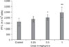
Due to the fact that the normal serum level of TNF-α was below the detection limit, we measured the TNF-α level of peritoneal macrophages stimulated by LPS. As shown in Fig. 9, the peritoneal macrophages TNF-α level was significantly increased in all BA-treated groups compared to the control group. In contrast, the serum concentration of IL-2 and IL-6 decreased significantly at the 0.5 mg/kg dose. No significant changes in the IL-10 serum level were observed in mice with or without BA treatment.
Discussion
In the current study, we clearly demonstrated that BA at various doses has a significant enhancement effect on cell-mediated immunity by promoting lymphocyte proliferation in response to immunological stimulation (via Con A or/and LPS) of T and B cells. Moreover, we found a BA-induced PFC increase in SRBC-immunized mice which indicates a positive effect of BA on humoral immune response.
Our observations were in accordance with other plant extracts; for example, extracts from Asparagus racemosus can activate murine splenocytes by markedly increasing their proliferation [14]. In the past few years, the percentage and ratio of two main lymphocyte T subsets, namely CD4+ cells or T helpers, and CD8+ or cytotoxic T cells, have been recognized as the most meaningful parameters for evaluating the balanced state of immunomodulation and response to homeostasis of the intrinsic immune system [9]. Previous studies have shown that some extractions from plants possess immune-stimulating capacity by increasing the percentage of CD4+ cells or CD4+/CD8+ ratio [4,14]. The present study provided similar a result showing significant increase in the percentage of CD4+ cells (Th cells) in thymus and the splenic ratios of CD4+/CD8+ in BA-treated groups. Furthermore, the percentage of CD19+ cells (total B cell) was increased in the spleen of mice exposed to BA. As a result, an increase in the relative number of T and B cells could possibly result in stimulating the immune response in BA-treated mice.
Currently, it is thought that CD4+ T cells differentiate into one of two effector phenotypes: T helper type 1 (Th1) cells which drive the immune response towards a cell-mediated immune response, and T helper type 2 (Th2) cells that promote a humoral or allergic response [8]. The functions of these subsets of T cells depend upon the specific types of cytokines that are generated, for example, IL-2, IFN-γ, and TNF-α by Th1 cells in contrast to IL-4, IL-5, IL-6, and IL-10 by Th2 cells. IL-10 is a negative immunoregulatory cytokine, which potentially suppresses Th1 functions such as the production of proinflammatory cytokines. These cytokines can dramatically affect not only the strength of the immune response, but also its character [21,29]. BA, an inhibitor of phospholipase A2, nitric oxide, and cyclooxygenase-2 [5,30] seems to be a modulator of cytokine production in Th1/Th2 cells. In our research, it was shown that BA significantly inhibited IL-2 (Th1) and slightly increased IL-10 (Th2) production. This observation suggests that BA has a mixed Th1 and Th2 adjuvant activity. However, its relative immunomodulatory effect turned out to be higher for Th2 as evidenced by a significant reduction in IL-2/IL-10 ratios, which is consistent with previous reports [30,31]. This data indicates that the compound stimulatory signals produced by immune cells decreased with the parallel increase in the inhibitory signals. On the hand, the serum level of IL-6 was decreased by BA. It is well-known that inappropriate regulation of IL-6 plays a protective as well as a deleterious role in antigen-specific immune-mediated diseases as characterized by low-grade inflammation [16,17]. IL-6 can also exert multiple stimulatory effects on cell growth and inflammation [26], and it is involved in the initiation and maintenance of the acute phase inflammatory response in immunoregulation and non-immune events in cells and tissues outside the immune system [11].
TNF-α is produced by Th1 cells, some Th2 cells, and cytotoxic T cells in both soluble and membrane-associated forms, and it can also deliver activating signals to macrophages. Some members of the TNF receptor family can stimulate apoptosis. Th2 cells express B cell-activating effector molecules, whereas Th1 cells express effector molecules that activate macrophages. We observed that TNF-α production was increased by BA in a dose-dependant manner (from 0.25 to 1 mg/kg). Thus, it may be hypothesized that BA is a modulator of cytokine production by Th1/Th2 subpopulations, but Th2 preferential adjuvant activity and its influence on inflammatory reactions in organisms may be very complex. The extracts from the bark of white birch (a plant known to be rich in BA) are often used in phytopharmacology as anti-cancer, anti-HIV, anti-bacterial, anti-inflammatory and anti-helminthic agents [31]. However, further studies are required for conclusive correlations.
Macrophages are not only the first line of defense in immune response to foreign invaders, but are also a kind of antigen-presentation cell which functions to present antigen-derived peptide fragments to T lymphocytes resulting in activating immunologically-competent T cells [3]. We found that BA also caused an increase in macrophage phagocytic activity. Some previous studies demonstrated that some agents of plant origin could enhance the capacity of the mononuclear phagocyte system resulting in stimulating host non-specific immunity [27,28]. Therefore, BA seems to enhance the antigen-presentation effect of macrophages by directly promoting phagocytosis capacity. Exposure to BA also caused an alteration in the serum levels of immunoglobulins. It is interesting to observe that BA was able to reduce the levels of IgM and IgG in mice. Since the concentration of immunoglobulins is associated with various pathological changes such as inflammation, activation of complement, and immune complex adhesion and immune deregulation, the decrease of IgM and IgG serum levels by BA can help attenuate these deleterious effects [4].
In conclusion, we have demonstrated that BA has the potential to enhance mouse immune function, including cellular immunity, humoral immunity, and activity of macrophages. Moreover, BA was a modulator of Th1/Th2 cells cytokine production, which suggests that this agent can be used for modulation of the immune system and as anti-inflammatory agent in animals. Further studies should be undertaken to determine the clinical significance of these findings, particularly in those with impaired immune function and/or in those with cancer.
 XML Download