

 PDF
PDF ePub
ePub Citation
 Print
Print


INTRODUCTION
Prostate-specific antigen (PSA) is the most widely used marker for the early detection of prostate cancer (PC) [1]. Patients who have elevated PSA levels or abnormal findings on a digital rectal examination (DRE) but whose biopsy results are negative pose a problem in PC screening. When the result of a DRE is normal and PSA is lower than 10 ng/mL, prostate biopsies fail to detect PC in 80% of men [2]. However, PSA is not a cancer-specific marker, and various benign processes also affect PSA concentrations [3,4]. Elevated PSA due to benign conditions (benign prostatic hyperplasia [BPH] and prostatitis) most directly underscores the difficulty in making a decision about repeat biopsy. The question remains as to the nature of the relationship between PSA and subclinical prostatic inflammation. Histological inflammation is a frequent finding in prostate biopsies that are performed on men without PC [5]. Several studies have investigated the relationship between PSA levels and the morphology of prostatic inflammation [6,7,8,9]. The results are controversial. Although it is known that acute prostatitis can contribute to lack of total PSA (tPSA) specificity, major disagreement remains over the effect of asymptomatic inflammation on free PSA (fPSA) and percentage of free PSA (f/tPSA) values [10,11,12,13]. It is essential to understand how factors other than PC, such as inflammation of the prostate, influence changes in PSA values. We therefore investigated the correlation between the morphological parameters of inflammation in prostate biopsies and tPSA, fPSA, and f/tPSA values in patients in the so-called "gray zone" (tPSA<10 ng/mL) and without clinically detectable PC.
MATERIALS AND METHODS
This study, which was performed from November 2008 to December 2012, included a series of 106 men with tPSA <10 ng/mL and/or f/tPSA<18% and who had undergone prostate biopsy that was negative for PC and showed no signs of prostatitis. Institutional Review Board approval was obtained before the initiation of the study. Serum PSA measurement (Immulite-DPC assay, Siemens, Erlangen, Germany), DRE, and transrectal ultrasound (Sonoline SI 400 U/S, Siemens) were performed on each patient. A biplanar transrectal probe was used with a frequency 5/7.5 MHz. Transrectal ultrasonography was used to determine the prostate volume [Vp=π/6× (D1×D2×D3)] and to guide the biopsies. Each patient underwent eight-core biopsy with an 18-gauge needle fitted to a Bard Magnum gun. Patients diagnosed with PC or premalignant lesions on biopsy were excluded from the study. Patients were also excluded from the study if they had a documented history or clinical signs of prostatitis, had PSA greater than l0 ng/mL, had prostate volume measured by transrectal ultrasound (TRUS) greater than 40 cm3, had previously undergone prostate surgery, had received 5-alpha-reductase inhibitors, or had an indwelling catheter or evidence of urinary tract infection (UTI).
Hematoxylin-eosin-stained slides taken from archival sections of biopsied prostatic tissue were analyzed by one pathologist. Each core of prostatic tissue (eight cores sampled from each patient) was scored for the type of inflammation and was graded by using a 4-point scale as follows: 0, no inflammatory cells (Fig. 1A); 1, mononuclear cell infiltrate (chronic inflammation; Fig. 1B); 2, mononuclear and polymorphonuclear cell infiltrate (chronic active inflammation; Fig. 1C); 3, polymorphonuclear cell infiltrate (acute inflammation; Fig. 1D). The effects of these morphologic aspects of inflammation were then correlated with serum tPSA, fPSA, and f/tPSA values.
In almost every set of biopsies, different types of histological inflammation were found. Thus, we used regression factor analysis to create a reduced classification for inflammation type. Factor analysis was applied according to the criteria of dimensionality reduction based on the number of derived variables. Input variables were the eight variables describing type of inflammation in individual biopsy cores of the prostate by use of the previously described 4-point scale. The criterion was to obtain a derived variable, that is, one factor on which each respondent was assigned a specific factor score. Instead of using four grades of inflammation for each biopsy core, we created two groups for statistical analysis. By use of regression analysis, each patient was given a unique inflammation type factor score (ITFS). The median of the factor scores was then calculated (ITFS median, 0.053). Patients with factor scores lower than or equal to the median (ITFS≤0.053) were included in the "more chronic inflammation" group. Similarly, those with factor scores higher than the median (ITFS>0.053) were included in the "more acute inflammation" group.
Descriptive statistics were used to characterize the age of patients, Vp, tPSA, fPSA, and f/tPSA. Comparisons of tPSA, fPSA, and f/tPSA values between groups were done by using Mann-Whitney U tests, and p<0.05 was considered statistically significant. Correlations among age, Vp, tPSA, fPSA, f/tPSA, and ITFS were determined by using Spearman's rank correlation analysis. For correlation analysis, p<0.01 was considered statistically significant. Data were analyzed by using R ver. 2.10.1 (Development Core Team. R: A language and environment for statistical computing. R Foundation for Statistical Computing, Vienna, Austria, 2009).
RESULTS
Data for 106 patients who met the inclusion criteria were analyzed. The median (range) age of the patients was 65 years (47 to 83 years) and the patients' median (range) prostate volume (Vp) was 31.5 cm3 (16 to 40 cm3). The median (range) levels of tPSA, fPSA, and f/tPSA were 6.6 ng/mL (3.4 to 10 ng/mL), 0.9 ng/ml (0.14 to 3.1 ng/mL), and 13.45% (2.5% to 35%). All patients had a Vp of less than 40 cm3 to minimize the effect of adenoma on PSA values.
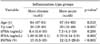
After initial histological coding of each biopsy core (eight cores per prostate biopsy) for inflammatory type, different types of histological inflammation were found in almost every set of prostate biopsies. Therefore, instead of having 4 grades of inflammation type for each biopsy core and for simplification purposes, we used regression analysis to create a reduced classification for inflammation type with two groups of patients for comparison: patients with more chronic inflammation (n=61, 57.5%) and patients with more acute inflammation (n=45, 42.5%).
The clinical characteristics and differences between the two groups according to grade of inflammation are shown in Table 1. The median (range) levels of tPSA, fPSA, and f/tPSA were 6.4 ng/mL (3.4 to 10 ng/mL), 1.09 ng/mL (0.28 to 3.1 ng/mL), and 15% (5.5% to 35%) for the chronic inflammation group, and 7.3 ng/mL (4.1 to 10 ng/mL), 0.79 ng/mL (0.14 to 2.9 ng/mL), and 12% (2.5% to 29.6%) for the acute inflammation group. A significant difference was found in fPSA (p=0.003) and f/tPSA (p<0.001) levels between groups, whereas the difference in tPSA levels was not significant (p=0.200). However, patients with more acute inflammation seemed to have slightly increased tPSA values. A positive correlation existed but was not significant (r=0.12, p=0.210). The fPSA and f/tPSA values were significantly decreased in the group of patients with more acute inflammation than in the patients with more chronic inflammation in the biopsy specimens.
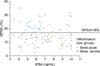
When we looked at the distribution of inflammation groups related to tPSA and f/tPSA values, patients with more acute inflammation tended to group below the horizontal line that represented the 18% f/tPSA cutoff for better discrimination between PC and benign conditions (Fig. 2). More acute histological inflammation decreased the percentage of free PSA, a tendency similar to that in PC.
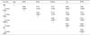
With the use of Spearman's analysis (Table 2), there was a significant negative correlation between type of inflammation and fPSA (r=-0.31, p=0.001) and f/tPSA (r=0.43, p<0.001) PSA values, in that a decrease in fPSA and f/tPSA values was found in patients with more acute inflammation. Total PSA values correlated significantly with fPSA (r=0.4, p<0.001) but not with type of inflammation (r=0.12, p>0.01). As expected, no correlation emerged between Vp and tPSA values (r=0.12, p=0.22), because only patients with small prostates (<40 cm3) were included in the study. On the other hand, Vp correlated with age (r=0.29, p=0.003), fPSA (r=0.32, p=0.001), and f/tPSA (r=0.31, p=0.001). Also, there was a significant negative correlation between Vp and inflammation type (r=-0.48, p<0.001).
DISCUSSION
We are often confronted with the association of abnormal PSA levels and biopsies that reveal no PC but only inflammation. Should the abnormal PSA levels be interpreted as a sign of missed PC, or can an inflammation explain the elevation of PSA? Although the determination of PSA is the best tool for the early detection of PC, the diagnostic accuracy of PSA is limited because of limited sensitivity and specificity, particularly in men with tPSA levels up to 10 ng/mL. The ratio of free-to-total PSA, calculated as percentage of fPSA, has been suggested as a useful tool for differentiating between PC and BPH, because the ratio is lower in PC than in BPH [14]. It is known that prostatitis can contribute to a rise in PSA, but disagreement exists about the effect of histological inflammation on PSA and its fractions. Recent results suggest that a decreased f/tPSA is not only characteristic of PC [15,16,17]. It is suggested that subclinical inflammation could affect both tPSA and fPSA values.
The best predictor of the serum PSA in patients with BPH but without PC is the size of the prostate [18]. Meyer at al. [13] reported that the serum PSA increased only in patients with benign prostates larger than 40 g. To minimize the effect of prostatic adenoma on PSA values, we included only patients with Vp up to 40 g. As expected, no correlation emerged between Vp and tPSA values (r=0.12, p=0.220). By contrast, we found a positive correlation between Vp and fPSA values (r=0.32, p=0.001). Similar results were reported by Scattoni et al. [19].
While some authors did not find any correlation between tPSA levels and subclinical inflammation, others found the correlation to be significant [9,20]. Okada et al. [17] reviewed cases of negative prostate biopsy results. In that study, the presence of inflammation correlated significantly with tPSA levels. Brawer et al. [6] found elevated tPSA levels only in patients with clinically acute prostatitis. Pansadoro et al. [7] found elevated tPSA values in 71% of patients with acute prostatitis, 15% of patients with chronic prostatitis, and 6% of patients with nonbacterial prostatitis. The results from our study agree with those of Nickel et al. [9] and Kwak et al. [8], who found no correlation between tPSA levels and type or extent of inflammation.
The present study was performed to draw attention to the effect of subclinical inflammation on fPSA and f/tPSA values. Our results confirm our hypothesis and those of others that more acute histological inflammation of the prostate induces a significant decrease of fPSA and f/tPSA values, compared with more chronic inflammation, although the scattergram of f/tPSA related to tPSA values showed overlap among all data (Fig. 2) [12,13]. Patients with more acute inflammation tended to group below the horizontal line that represented the 18% f/tPSA cutoff for more efficient discrimination between PC and benign conditions. That cutoff was previously shown to be the limit with the highest efficiency to differentiate between BPH and PC [21]. These data emphasize that all benign prostatic diseases do not differ from PC in regard to f/tPSA.
Only a few studies have evaluated fPSA in patients with asymptomatic inflammation. Many studies confirm that it can cause a rise of tPSA, but there are major discrepancies in findings when we look at fPSA values. Morote et al. [11] studied patients without evidence of PC in TRUS-guided biopsies. In that study, the size of the prostate was the only significant contributor to the PSA concentration. In a study performed by Minardi et al. [22], inflammation associated with BPH compared with BPH only led to false-positive tPSA and f/tPSA levels, because 60% of these patients had an f/tPSA ratio below l6%. Our results comply with those of Jung et al. [12] and Meyer et al. [13], who reported reduced f/tPSA values in patients with PC and inflammation compared with controls or BPH patients. On the other hand, our results differ from those of Ornstein et al. [10], who showed that f/tPSA levels do not appear to be altered by inflammation. However, those results must be considered preliminary because Ornstein et al. [10] did not stratify the patients by the type of inflammation present. Opposite to our results, Scattoni et al. [19] reported that histologically more acute inflammation induces an increase in the f/tPSA ratio, whereas chronic inflammation seems to induce lower f/tPSA ratios in patients without PC. Because our results confirm that histological inflammation (especially more acute) is characterized by slightly elevated tPSA (and decreased fPSA values), f/tPSA fails to differentiate between cancerous and noncancerous prostatic disease.
It is assumed that inflammation increases PSA concentrations by causing leakage of PSA from acini. However, increased PSA concentrations have only been found when glandular epithelium was disrupted [23]. Recent studies have demonstrated that acute, clinical inflammation produces a more intense or different process of PSA release than does subclinical inflammation of the prostate [24,25]. All our patients had different patterns of inflammation but had no signs of clinical prostatitis. Changes in tPSA values between groups were minimal and were not statistically significant (the acute inflammation group had slightly higher tPSA values), but patients with more acute histological inflammation had significantly decreased fPSA and f/tPSA values. The molecular reasons for these inflammation-related changes in the PSA forms are still not defined. PSA exists in different forms [26]. It is predominantly bound to αl-antichymotrypsin with approximately 70% to 90% of tPSA in serum. The approximately 10% to 30% of tPSA that is not bound to proteins represents fPSA. Thus, changes in f/tPSA reflect changes in fPSA or PSA α1-antichymotripsin. As in PC, the changed f/tPSA in subclinical prostatitis similarly proves that PSA α1-antichymotripsin is elevated compared with fPSA. Bjork et al. [26] showed that α1-antichymotripsin is also synthesized in PC cells but not in BPH cells. In that way, more PSA is complexed in PC than in BPH, leading to the changes observed in serum. Similar changes can occur in prostatic inflammation, because increased α1-antichymotripsin production is a typical reaction to inflammation. Another explanation was proposed by Zackrisson et al. [27], who studied the evolution of tPSA and fPSA and their ratios during 1-year follow-up after febrile UTI [27]. Most men in their study had a rise in fPSA and tPSA during the acute phase of the UTI. After 1 month, fPSA rapidly decreased to the levels that were maintained during the rest of the follow-up period, but elevations of tPSA were sustained for up to 6 months after the acute phase of UTI. These changes in tPSA and fPSA levels during the chronic phase of UTI could falsely be interpreted as a sign of PC. It was previously shown that the fPSA molecule has a much shorter mean half-life than does tPSA [26].
Our results imply that the decreased level of serum fPSA and f/tPSA is not typical of PC alone, because these values are similarly affected by subclinical inflammation. Despite the significant effect of subclinical inflammation on fPSA and f/tPSA levels, we cannot deduce if the f/tPSA is a reliable discriminator between PC and prostatitis. Neither clinical examination nor PSA permits reliable discrimination of PC and prostatitis, especially in patients with PSA levels up to 10 ng/mL. These patients should undergo prostate biopsy. On the other hand, when we find considerable inflammation in our biopsy report, but no evidence of PC, long-term antibiotic or anti-inflammatory therapy seems like a reasonable option. Bozeman et al. [28] showed that the use of antibiotics for 4 weeks normalized tPSA and fPSA levels in 30% of studied patients. In this way, unnecessary biopsies could be avoided in a considerable number of patients.
CONCLUSIONS
Inflammation has an important prevalence in cancer-free prostate biopsy specimens. Subclinical inflammation seems to have a significant influence on fPSA and f/tPSA values in patients with tPSA levels up to 10 ng/mL but without detectable PC. Subclinical inflammation is not characterized by elevated tPSA concentrations alone but also by a decreased percentage of fPSA, a tendency similar to that in PC. In the assessment of biopsy results negative for PC, it might be helpful to evaluate the degree and extension of histological inflammation, especially in terms of the necessity for subsequent repeated biopsies.

 XML Download