

 PDF
PDF ePub
ePub Citation
 Print
Print


Abstract
Androgen deprivation therapy, which is the standard treatment for metastatic prostate cancer, includes nonsteroidal antiandrogenic drugs, such as flutamide, nilutamide and bicalutamide. Of them, bicalutamide rarely induces interstitial pneumonia. We report a case of bicalutamide-induced interstitial pneumonia. A 68-year old male diagnosed with prostate cancer and multiple bone metastases presented with dry cough and low grade fever for 3 days. He had taken bicalutamide (50 mg/day) for 13 months. High resolution computed tomography revealed ground glass opacity in his right upper lung. The laboratory studies showed no eosinophilia in the serum and bronchoalveolar lavage fluid. Despite the use of antimicrobial agents for 2 weeks, the extent of the lung lesions increased to the left upper and right lower lung. He had no environmental exposure, collagen vascular disease and microbiological causes. Under the suspicion of bicalutamide-induced interstitial pneumonia, bicalutamide was stopped and prednisolone (1 mg/kg/day) was initiated. The symptoms and radiologic abnormalities were resolved with residual minimal fibrosis.
Figures and Tables
Figure 1
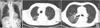
(A) Chest radiography shows interstitial pneumonia in right upper lobe at the presentation. (B) Chest CT shows consolidation and bronchiectasis with cystic formation in right upper lobe at the presentation.
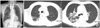
Figure 2
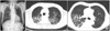
(A) Interstitial pneumonia extends to left upper lobe, compared with chest radiography at the presentation. (B) Chest CT shows newly developed diffuse interlobular septal thickening and ground glass attenuation with bronchiectasis in left upper lobe.
References
1. Camus P, Fanton A, Bonniaud P, Camus C, Foucher P. Interstitial lung disease induced by drugs and radiation. Respiration. 2004. 71:301–326.
2. Pfitzenmeyer P, Foucher P, Piard F, Coudert B, Braud ML, Gabez P, et al. Nilutamide pneumonitis: a report on eight patients. Thorax. 1992. 47:622–627.
3. Azuma T, Kurimoto S, Mikami K, Oshi M. Interstitial pneumonitis related to leuprorelin acetate and flutamide. J Urol. 1999. 161:221.
4. Shioi K, Sakai N, Yoshida M, Nakamura M. Successful recovery from interstitial pneumonitis, induced by bicalutamide and leuprorelin acetate given as treatment for prostate cancer. Hinyokika Kiyo. 2005. 51:211–214.
5. Shioi K, Yoshida M, Sakai N. Interstitial pneumonitis induced by bicalutamide and leuprorelin acetate for prostate cancer. Int J Urol. 2003. 10:625–626.
6. Mayaud C, Fartoukh M, Parrot A, Cadranel J, Milleron B, Akoun G. Drug-associated interstitial lung disease: a diagnostic challenge. Rev Pneumol Clin. 2005. 61:179–185.
7. Ben-Noun L. Drug-induced respiratory disorders: incidence, prevention and management. Drug Saf. 2000. 23:143–164.
8. Ellis SJ, Cleverley JR, Muller NL. Drug-induced lung disease: high-resolution CT findings. AJR Am J Roentgenol. 2000. 175:1019–1024.
9. Landis SH, Murray T, Bolden S, Wingo PA. Cancer statistics, 1999. CA Cancer J Clin. 1999. 49:8–31.
10. Dahele M, Brade A, Pearson S, Bezjak A. Stereotactic radiation therapy for inoperable, early-stage non-small-cell lung cancer. CMAJ. 2009. 180:1326–1328.
11. Kim TH, Cho KH, Pyo HR, Lee JS, Zo JI, Lee DH, et al. Dose-volumetric parameters for predicting severe radiation pneumonitis after three-dimensional conformal radiation therapy for lung cancer. Radiology. 2005. 235:208–215.
12. Koenig TR, Munden RF, Erasmus JJ, Sabloff BS, Gladish GW, Komaki R, et al. Radiation injury of the lung after three-dimensional conformal radiation therapy. AJR Am J Roentgenol. 2002. 178:1383–1388.
13. Ikezoe J, Takashima S, Morimoto S, Kadowaki K, Takeuchi N, Yamamoto T, et al. CT appearance of acute radiation-induced injury in the lung. AJR Am J Roentgenol. 1988. 150:765–770.
14. Cleverley JR, Screaton NJ, Hiorns MP, Flint JD, Muller NL. Drug-induced lung disease: high-resolution CT and histological findings. Clin Radiol. 2002. 57:292–299.
 XML Download