

 PDF
PDF ePub
ePub Citation
 Print
Print


INTRODUCTION
Lung cancer is a common tumor malignancy with high mortality worldwide.1 And lung adenocarcinoma (LA) is a major subtype that accounts for 40% of lung cancer with poor prognosis.2 With the development of chemo-therapy and immunological therapy, more strategies have been implicated in treatment of LA, whereas the survival rate is still low.3 Emerging research have reported that immune system is responsible for immunotherapy of lung cancer.4 Natural killer (NK) cell is one of the key immune cells and its cytolytic potential is limited in lung cancer, whereas increasing NK cells function can result in tumor regression.5 Potentiation of cytotoxicity, mediated by NK cells to LA, plays a vital role in overcoming tumor cell escape from immune system.6 NK cells control tumor by regulating pro-inflammatory cytokines production, such as interferon-γ (IFN-γ) and tumor necrosis factor-α (TNF-α), which can induce cytolysis of tumor cells.7 Therefore, it is promising to explore a novel driver that modulates the killing effect of NK cells to LA cells.
MicroRNAs (miRNAs) hold great promise in development of LA, and are associated with prognosis of LA.8 Previous research have reported that miRNAs may regulate the function of NK cells by enhancing or inhibiting the cytotoxic potential of NK cells.9 Moreover, miRNAs have been suggested to participate in NK cells activity impairment.10 For example, miR-183 is associated with NK cells function in lung cancer.11 The available evidence indicates that miR-218-5p plays an essential role in LA progression by regulating target gene expression.12 Notably, miR-218-5p is expressed in human NK cells, and may be involved in the development and function of NK cells.13 Therefore, we speculate miR-218-5p may be implicated in regulating NK cells function in LA.
A number of researchers have reported that serine hydroxymethyl transferase 1 (SHMT1) is required for tumor growth and disease progression.1415 Previous work investigated the regulation of SHMT1 on lung cancer risk, and suggested SHMT1 may affect etiology of lung cancer.16 In LA, SHMT1 has an important impact on cell proliferation and apoptosis.17 However, there is no direct evidence in support of SHMT1 required for NK cells function. Intriguingly, bioinformatics analysis predicates the putative binding sites of miR-218-5p and SHMT1. Therefore, we assumed that SHMT1 is required for miR-218-5p-mediated function on the killing effect of NK cells to LA cells. In the present study, we detected the expressions of miR-218-5p and SHMT1 in NK cells of LA patients, and explored the potential mechanism that underlies miR-218-5p regulating the killing effect of NK cells to LA cells.
MATERIALS AND METHODS
Isolation of primary NK cells
Peripheral blood mononuclear cells were collected from 20 patients with LA and healthy volunteers, and were used to isolate human NK cells using NK cell enrichment kit (Thermo Fisher, Wilmington, DE, USA) according to the manufacturer's instructions. Highly pure NK cells were obtained by negative selection with magnetic labeling. Informed consent was signed by all patients, and the study was accepted by the Institutional Research Ethics Committee of The Affiliated Renhe Hospital of China Three Gorges University.
Cell culture and treatment
Human NK cell line NK-92, mouse NK cell line LNK, human LA cell line A549, and 293T cells were obtained from American Tissue Culture Collection (ATCC, Manassas, VA, USA). In this study, all cells were grown in RPMI-1640 (Gibco, Carlsbad, CA, USA) containing 10% fetal bovine serum (Gibco), 1% penicillin, and streptomycin (Invitrogen, Carlsbad, CA, USA) at 37℃ in an incubator with 5% CO2.
To activate NK-92 or LNK cells, 20 ng/mL interleukin-2 (IL-2, Gibco) was introduced into cells for 24 h. SHMT1 overexpression vectors (SHMT1) cloned into pcDNA, miR-218-5p mimics (miR-218-5p), miR-218-5p inhibitor (anti-miR-218-5p), and negative control (miR-NC or anti-NC) were obtained from GenePharma (Shanghai, China). Transfection was performed in NK-92 or LNK cells using Lipofectamine 2000 (Invitrogen) according to the manufacturer's protocol.
Quantitative real-time polymerase chain reaction
Total RNA was isolated using Trizol reagent (Invitrogen) according to the manufacturer's instructions. Then cDNA was synthesized using TaqMan miRNA reverse transcription kit (Applied Biosystems, Foster City, CA, USA), followed by quantitative real-time polymerase chain reaction (qRT-PCR) with SYBR Green Master Mix (Applied Biosystems). All primers were listed as follows: miR-218-5p (Forward, 5′-TTGCGGATG GTTCCGTCAAGCA-3′; Reverse, 5′-ATCCAGTGCAGGGTCC GAGG-3′), U6 (Forward, 5′-CTCGCTTCG GCAGCACA-3′; Reverse, 5′-AACGCTTCACGAATTTGCGT-3′), SHMT1 (Forward, 5′-TTGCCTCGGAGAATTTCGCC-3′; Reverse, 5′-GTCCCGC CATAGT ATCTCTGG-3′), and β-actin (Forward, 5′-CAGCCTT CCTTCTTGGGTAT-3′; Reverse, 5′-TGGCATAGAGGTCTT TACGG-3′). The expression of miR-218-5p or SHMT1 was analyzed using 2−ΔΔCt method, and normalized to U6 or β-actin, respectively.
Western blots
Total protein was prepared in cell lysis buffer containing 1% protease inhibitor (Thermo Fisher). After quantification by BCA assay kit (Sigma, St. Louis, MO, USA), denatured proteins were separated by SDS-PAGE gel, transferred to polyvinylidene difluoride membranes (Millipore, Billerica, MA, USA), and then blocked with blocking reagent (Thermo Fisher) for 1 h at room temperature. Subsequently, the membranes were incubated with primary antibodies against SHMT1 or β-actin (Cell Signaling Technology, Danvers, MA, USA) overnight at 4℃. After being rinsed with TBST, membranes were hatched with secondary antibodies (Cell Signaling Technology) for 2 h at room temperature. The protein blots were visualized using enhanced chemiluminescence chromogenic substrate (Thermo Fisher), and investigated by Image Lab software (Bio-Rad, Hercules, CA, USA). β-actin was used as a standard for band intensities.
Enzyme linked immunosorbent assay
After treatment, cell supernatants were collected to detect levels of IFN-γ and TNF-α using commercial human ELISA Kit (Invitrogen) according to the manufacturer's instructions. Intensity of color was assayed at 450 nm with reference wave length at 620 nm using a microplate reader (Bio-Rad).
Cytotoxicity assay
The cytotoxicity of NK-92 to A549 cells was investigated by a lactate dehydrogenase (LDH) assay according to the manufacturer's instructions. Briefly, A549 cells were seeded into 96-well plates at a density of 1×10⁴ cells per well, and then IL-2-treated NK-92 cells transfected with miR-218-5p, miR-NC, anti-miR-218-5p, anti-NC, pcDNA, or SHMT1 were introduced into each well, and co-cultured with A549 cells at effector/target cell ratio of 5:1 for 4 h. Following incubation, supernatants were collected, and the cytotoxicity of NK-92 cells was detected by LDH cytotoxicity assay kit (Thermo Fisher).
Luciferase activity assay
TargetScan analysis predicted putative binding sites of miR-218-5p and 3′ untranslated regions (3′-UTR) sequences of SHMT1. Luciferase report vectors with wild-type plasmid (SHMT1-wt) or mutant-type plasmid (SHMT1-mut) were generated using pGL3 vector (Promega, Madison, WI, USA), respectively. Luciferase reporter vectors, miR-218-5p, anti-miR-218-5p, or their negative control were co-transfected in 293T cells for 48 h using Lipofectamine 2000 according to the manufacturer's protocols. Luciferase activity was evaluated by luciferase assay kit (GeneCopoeia, Rockville, MD, USA).
Animals and xenograft model of LA
Every protocol was approved by the Animal Research committee of The Affiliated Renhe Hospital of China Three Gorges University. SPF BALB/c nude mice (male, 6-week-old) were purchased from Vital River Laboratory Animal Technology (Beijing, China). A549 cells (6×10⁶ cells) were introduced into nude mice by subcutaneous injection (n=8 per group). At 2 h after LA model construction, IL-2-treated LNK cells (3×10⁶ cells), with stable transfection of anti-miR-218-5p or anti-NC, were subcutaneously injected into the same sites. Tumors were examined every three days, and tumor volume was calculated with slide calipers by (0.5×length×width×height). Mice were sacrificed at 15 days after cell implantation, and tumor specimens were collected for molecular analyses.
RESULTS
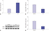
MiR-218-5p was enhanced while SHMT1 expression was impaired in NK cells of LA patients
To test whether miR-218-5p and SHMT1 participate in functioning of NK cells, their expressions were detected by qRT-PCR and western blots in NK cells from healthy control and LA patients (n=20). As a result, miR-218-5p expression was significantly increased in NK cells of LA patients compared to control group (Fig. 1A). However, a strong reduction of SHMT1 mRNA level was observed in NK cells in LA group compared to control group (Fig. 1B). Moreover, SHMT1 protein abundance showed similar trend in NK cells (Fig. 1C and D). These finding indicated that miR-218-5p and SHMT1 might play essential roles in NK cells in LA.
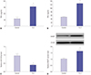
Activated NK-92 cells promoted cytokines production, inhibited miR-218-5p, and induced SHMT1 protein expression
IL-2 was used to induce activation of NK cells in vitro. NK-92 cells showed enhanced TNF-α production after IL-2 stimulation (Fig. 2A). Meanwhile, IFN-γ expression was also elevated in NK-92 cells in response to IL-2 (Fig. 2B). These suggested that NK cells were activated by IL-2 treatment, indicating triggering of killing effect of NK cells. Subsequently, the expressions of miR-218-5p and SHMT1 were detected in activated NK-92 cells. The expression of miR-218-5p was impaired in activated NK cells compared to those without IL-2 treatment (Fig. 2C). Besides, IL-2 insult resulted in a great increase of SHMT1 protein level in NK cells compared to that before IL-2 stimulation (Fig. 2D). These results further showed that miR-218-5p and SHMT1 might be required for the functioning of NK cells.
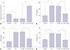
Addition of miR-218-5p blocked the killing effect of NK cells to LA cells
To evaluate the effect of miR-218-5p on killing effect of NK cells to LA cells, miR-218-5p mimics were introduced into NK-92 cells and co-cultured with A549 cells after IL-2 stimulation. MiR-218-5p expression was downregulated in NK-92 cells after IL-2 treatment, whereas addition of miR-218-5p reversed the level of miR-218-5p (Fig. 3A). Moreover, abundant presence of miR-218-5p inhibited IFN-γ and TNF-α secretion in NK-92 cells with treatment of IL-2, which activated cells and promoted cytokines production (Fig. 3B and C). In addition, the cytotoxicity of activated NK cells to A549 cells was investigated by co-culturing of NK-92 and A549 cells. Results showed that IL-2 stimulation improved the killing effect of NK-92 cells, while introduction of miR-218-5p displayed opposite effect (Fig. 3D). These results suggested that miR-218-5p overexpression can protect against the killing effect of NK cells to LA cells.
SHMT1 was a target of miR-218-5p
Since miR-218-5p and SHMT1 were involved in dysfunction of NK cells, we next probed the potential link of miR-218-5p and SHMT1 in NK-92 cells. Bioinformatics assay described the putative binding sites of miR-218-5p and SHMT1 by TargetScan, indicating SHMT1 might be a direct target of miR-218-5p (Fig. 4A). Next, we investigated the interaction in 293T cells by luciferase activity assay. SHMT1-wt and miR-218-5p were co-transfected into 293T cells, and luciferase activity was obviously inhibited in miR-218-5p group compared to NC group, whereas there was not significant difference of the activity in response to SHMT1-mut (Fig. 4B). Conversely, luciferase activity of cells with SHMT1-wt showed a higher level in anti-miR-218-5p group compared to that in anti-NC group. However, miR-218-5p deletion failed to show efficacy in affecting the activity of cells transfected with SHMT1-mut (Fig. 4C). These uncovered the interaction between miR-218-5p and SHMT1. Accordingly, the effect of miR-218-5p on SHMT1 expression was investigated in NK-92 cells. Addition of miR-218-5p led to a great loss of SHMT1 mRNA and protein level compared to NC group (Fig. 4D and E). Nevertheless, the absence of miR-218-5p showed an opposite effect, revealed by elevated SHMT1 abundance at mRNA and protein level (Fig. 4F and G), indicating miR-218-5p was negatively correlated with SHMT1 in NK cells.
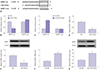
SHMT1 was required for miR-218-5p-mediated killing effect of NK cells to LA cells
To probe whether SHMT1 was associated with miR-218-5p-mediated limited capacity for killing effect of NK cells, miR-218-5p or (and) SHMT1 were transfected into IL-2-treated NK-92 cells. MiR-218-5p abundance was markedly impaired after IL-2 insult and miR-218-5p mimics protected the expression, while SHMT1 overexpression could not afford miRNA expression (Fig. 5A). Besides, SHMT1 expression was promoted in NK-92 cells by IL-2 stimulation and miR-218-5p addition inhibited SHMT1 abundance, whereas SHMT1 overexpression effectively increased protein levels (Fig. 5B). IL-2 facilitated IFN-γ production and miR-218-5p blocked cytokine secretion, while SHMT1 reversed the expression of IFN-γ in NK-92 cells (Fig. 5C). Moreover, similar trend was uncovered in terms of TNF-α level in NK cells (Fig. 5D). Likewise, SHMT1 ablated the effect of miR-218-5p on cytotoxicity of NK cells to A549 cells, which enhanced the killing effect (Fig. 5E). These data indicated that miR-218-5p overexpression repressed the killing effect of NK cells to LA cells by regulating SHMT1 expression.
MiR-218-5p depletion limited LA tumor growth in vivo
To further explore the effect of miR-218-5p on cytotoxicity of NK cells to LA cells in vivo, activated LNK cells were transfected with anti-miR-218-5p or anti-NC, and then introduced into mice model of LA (n=8 per group). Tumor volume was progressively increased, and absence of miR-218-5p decreased tumor growth compared to anti-NC treatment group (Fig. 6A). Moreover, the expression of SHMT1 was examined in tumor tissues. An evident increase in protein level was observed in miR-218-5p deficiency group compared to NC group (Fig. 6B and C). These findings suggested that miR-218-5p exhaustion weakened tumor growth in vivo by enhancing the killing effect of NK cells to LA cells.
DISCUSSION
LA is a major subtype of lung cancer which threatens people's health all over the world.2 Over the past few years, immune-based therapy has gained more attention, and increasing number of investigators showed great promise in cytotoxicity of NK cells to tumor cells. NK cells played important roles in various types of cancer, including hepatocellular carcinoma and colorectal cancer.1819 Likewise, immune T cell and NK cell were involved in LA progression by varying pathways.20 Besides, miRNAs have been suggested to be associated with control of immune response in diverse cancers by regulating functions of T or NK cells.21 For instance, miR-146a suppressed NK cells function via mediating signal transducer and activator of transcription 1 (STAT1).22 However, miR-30c could confer NK cells cytotoxicity by regulating natural-killer group 2 member D (NKG2D).23 A novel miRNA, miR-218-5p, has been reported as a potential biomarker in many cancers, such as colorectal cancer, osteoarthritis, and LA.242526 However, the study on involvement of miR-218-5p in NK cells function remains poorly understood. In the present study, we obtained NK cells from LA patients or healthy controls, and detected the expressions of miR-218-5p and SHMT1 in NK cells, and showed that both miR-218-5p and SHMT1 were expressed in NK cells.
Tumor cells have been reported to be killed by immune cells, while tumor immune escape supported tumor development by triggering the resistance to cytotoxicity of immune cells, such as NK cells.5 Previous research revealed that transforming growth factor-β (TGF-β) and IL in the microenvironment might disturb the balance of limitation of NK cell function in cancers.27 Furthermore, IL-2 was widely used to induce activation of NK cells to inhibit tumor processes.2829 Therefore, we also introduced IL-2 into NK cells and found that IL-2 stimulated IFN-γ and TNF-α secretion, and altered the abundances of miR-218-5p and SHMT1. This was consistent with another finding that suggested miR-218-5p had low expressions in activated NK cells.13 We hypothesized that miR-218-5p and SHMT1 might be required for activation of NK cells, which could provide a novel biomarker of regulating NK cells function. To test the hypothesis, we probed whether miR-218-5p might affect the killing effect of NC cells to LA cells. Our results showed that miR-218-5p overexpression inhibited inflammatory cytokines production and cytotoxicity against LA cells, indicating that miR-218-5p was negatively correlated with the killing effect of NK cells. Similarly, miR-889 compromised the killing effect of NK cells by increasing resistance to cytotoxicity of NK cells.18 Apart from miR-889, miR-615-5p also blocked NK cells cytotoxicity to tumor cells by regulating insulin-like growth factor-1R (IGF-1R) expression.30 These findings uncovered that miRNAs might play an essential role in regulating NK cells cytotoxicity.
miRNAs interacted with the development and function of NK cells by transcriptional and post-transcriptional regulation.31 MiR-218-5p was negatively correlated with poor prognosis of lung cancer by targeting IL-6 and STAT3.32 Besides, slug and zinc finger E-box binding homeobox 2 (ZEB2) were also targeted by miR-218-5p, and played essential roles in epithelial-mesenchymal transition and metastasis in lung cancer.33 SHMT1 was reported to be crucial for tumor progression in various cancer types, including lung cancer.141634 In addition, SHMT1 might participate in cell proliferation and apoptosis by miR-198 targeting in LA.17 Previous study revealed that vitamin B6 might regulate inflammatory response by mediating SHMT-related pathway.35 Moreover, SHMT1 was suggested to stimulate the production of inflammatory cytokines in ovarian cancer.36 Besides, currently available evidence have indicated SHMT1 was required for B-lymphoma cell growth.34 These findings uncovered that SHMT1 might be regarded as a target for immunotherapy of tumors. However, the mechanism that SHMT1 requires for functioning of NK cells still remains unclear. Hence, we probed a link of miR-218-5p with SHMT1, and investigated the effect of SHMT1 on functioning of NK cells. Our results showed that addition of SHMT1 ameliorated NK cell function. Murine xenograft model in vivo was responsible for preclinical drug trials, and it has been widely used for NK cells function or LA progression investigation.2937 In this study, we found that miR-218-5p also regulated the killing effect of NK cells in vivo, revealed by reduced tumor volume. MiR-218-5p and SHMT1 were involved in not only cytotoxicity of NK cells but also in LA progression, whereupon the mechanism by which miR-218-5p and SHMT1 affected LA processes should be investigated further in future studies.
In summary, miR-218-5p was enhanced in NK cells but inhibited by IL-2 treatment. Moreover, miR-218-5p overturned the killing effect of NK cells to LA cells by regulating SHMT1 expression in vitro and in vivo. This might indicate that miR-218-5p has a potential to regulate NK cell functions to address anti-tumor role in cancers, such as LA.

 XML Download